. 2024 Aug 16;25(16):8941.
doi: 10.3390/ijms25168941.

Prolactin Drives Iron Release from Macrophages and Uptake in Mammary Cancer Cells through CD44

Affiliations

Prolactin Drives Iron Release from Macrophages and Uptake in Mammary Cancer Cells through CD44

Reagan Farrell et al. Int J Mol Sci. .

Abstract

Iron is an essential element for human health. In humans, dysregulated iron homeostasis can result in a variety of disorders and the development of cancers. Enhanced uptake, redistribution, and retention of iron in cancer cells have been suggested as an "iron addiction" pattern in cancer cells. This increased iron in cancer cells positively correlates with rapid tumor growth and the epithelial-to-mesenchymal transition, which forms the basis for tumor metastasis. However, the source of iron and the mechanisms cancer cells adopt to actively acquire iron is not well understood. In the present study, we report, for the first time, that the peptide hormone, prolactin, exhibits a novel function in regulating iron distribution, on top of its well-known pro-lactating role. When stimulated by prolactin, breast cancer cells increase CD44, a surface receptor mediating the endocytosis of hyaluronate-bound iron, resulting in the accumulation of iron in cancer cells. In contrast, macrophages, when treated by prolactin, express more ferroportin, the only iron exporter in cells, giving rise to net iron output. Interestingly, when co-culturing macrophages with pre-stained labile iron pools and cancer cells without any iron staining, in an iron free condition, we demonstrate direct iron flow from macrophages to cancer cells. As macrophages are one of the major iron-storage cells and it is known that macrophages infiltrate tumors and facilitate their progression, our work therefore presents a novel regulatory role of prolactin to drive iron flow, which provides new information on fine-tuning immune responses in tumor microenvironment and could potentially benefit the development of novel therapeutics.

Keywords: CD44 upregulation; iron transfer; macrophages; mammary cancer cells; prolactin.


Conflict of interest statement

The authors declare no conflicts of interest.

Figures

Figure 1
Figure 1
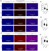
Regulation of intracellular labile iron pool by prolactin in mouse breast cancer cells under normal culture condition. Effect of prolactin treatment on the intracellular labile iron pool in EO771 cells (A) and Py230 cells (C). Blue and red fluorescence stains of the nucleus and labile iron pool, respectively. Quantification of the intracellular labile iron fluorescent intensity by prolactin treatment in EO771 cells (B) and Py230 cells (D). Results are calculated as the average labile iron fluorescence of all individual cell from three independent trials (n = 3). Data represent mean ± SD. (***, p < 0.001; ****, p < 0.0001).
Figure 2
Figure 2
Effects of prolactin treatment on gene expression involved in iron transport in E0771 cells. Changes in expression levels in the Fe3+ transporter TFRC (A), Fe2+ transporter DMT1 (B), Fe3+ and Fe2+ transporters including the hyaluronate receptor CD44 (C) and scavenger receptor CD163 (D), iron exporter FPN I (E), the negative regulator hepcidin for iron exporter (F), and the ratio of hepcidin to FPN I (G). Data represent mean ± SEM from three biological replicates (n = 3) (**, p < 0.01; n.s. = not significant). Comparison of iron transport-related gene expression between normal and breast cancer cell lines (H). The green and blue rectangles indicate downregulation and upregulation of the CD44 gene, respectively. Analysis of 2326 human patients with invasive breast carcinoma revealed significant types of CD44 mutations (x-axis) and its overexpression (y-axis) (I). Among these patients, a positive correlation (Spearman coefficient = 0.05, p = 0.04) was found between tumoral expression of CD44 (y-axis) and prolactin (x-axis) (J).
Figure 3
Figure 3
The upregulated CD44 by prolactin stimulation in EO771 cells contributed to iron uptake. (A) Representative image of live detection of CD44 expression in cultured EO771 cells over a two-day time course. Green fluorescence indicates the expression of CD44 in EO771 cells. (B) Quantification of green fluorescent intensity of CD44 expression per EO771 cell. Data represent mean ± SEM from twelve biological replicates (n = 12) (*, p< 0.05) (C) Percentage of CD44 expressing EO771 cells over two-day time course. Data represent mean ± SEM from twelve biological replicates (n = 12). Representative image ((D), control (E), and Prolactin treatment) and fluorescent quantification of intracellular labile iron pool through blockage of CD44 (F). Data represent mean ± SD. (**, p < 0.01; ****, p < 0.0001). Blue and red fluorescence stain represent the nucleus and labile iron pool, respectively.
Figure 4
Figure 4
Effects of prolactin treatment on gene expression involved in iron transport in macrophage cell line, RAW264.7. Changes in expression levels in the ferric iron transporter TFRC (A), ferrous iron transporter DMT1 (B), hyaluronate receptor CD44 (C), scavenger receptor CD163 (D), iron exporter FPN I (E), the negative regulator hepcidin for iron exporter (F), and the ratio of hepcidin to FPN I (G). The heatmap displays the expression of six genes related to macrophage polarization in response to prolactin treatment, including two M1 markers (NOS2 and TNFα) and four M2 markers (Arg2, CD163, IL10, and CD206) (H). Data represent mean ± SEM from three biological replicates (n = 3). (*, p < 0.05; **, p < 0.01; n.s. = not significant) Data represent the relative expressed transcripts of examined iron transport related genes compared to the housekeeping gene, TATA-box binding protein.
Figure 5
Figure 5
Regulation of intracellular labile iron pool by prolactin in macrophage cells, RAW264.7, under normal culture condition or extra-iron supplementations. (AC) Effect of prolactin treatment on the intracellular labile iron pool in RAW264.7 under normal culture condition (A), supplemented with extra ferrous iron (reduced state) (B), or supplemented with extra ferric iron (oxidized state) (C). Blue and red fluorescence stains represent the nucleus and labile iron pool, respectively. (DF) Quantification of the intracellular labile iron fluorescent intensity by prolactin treatment in RAW264.7 cells under normal (D), extra ferrous (E), or extra ferric iron supplementation (F). Results are calculated as the average labile iron fluorescence of all individual cells from three independent trials (n = 3). Data represent mean ± SD. (****, p < 0.0001).
Figure 6
Figure 6
Direct iron transfer from co-cultured macrophages to breast cancer cells. (A) Iron transfer occurs in a very short time frame (4 h) between Py230 and RAW264.7 cells. (B) Iron transfer occurs in a very short time frame (2 h) between EO771 and RAW264.7 cells. Representative images are selected from three biological replicates. Blue, green, and red fluorescence stains represent nuclei, cytoplasm, and labile iron pools, respectively.
Figure 7
Figure 7
Flow cytometric analysis validates the direct iron transfer from macrophages to breast cancer cells. No fluorescent iron signal was found in EO771 cells when not co-cultured with macrophages (A) but iron fluorescence was seen when co-cultured with either control macrophages (B) or macrophages pretreated with prolactin (C). (D) More EO771 cells received fluorescent iron from prolactin pre-treated macrophages. Data represent mean ± SD from five biological replicates (n = 5). (*, p < 0.05). Staining controls for flow cytometry are shown in (E) (unstained macrophages), (F) (iron and MHC II stained macrophages), (G) (unstained EO771 cells), and (H) (iron and MHC II stained EO771 cells).

References

    1. Ganz T. Systemic iron homeostasis. Physiol. Rev. 2013;93:1721–1741. doi: 10.1152/physrev.00008.2013. - DOI - PubMed
    1. Das B.K., Wang L., Fujiwara T., Zhou J., Aykin-Burns N., Krager K.J., Lan R., Mackintosh S.G., Edmondson R., Jennings M.L., et al. Transferrin receptor 1-mediated iron uptake regulates bone mass in mice via osteoclast mitochondria and cytoskeleton. elife. 2022;11:e73539. doi: 10.7554/eLife.73539. - DOI - PMC - PubMed
    1. Bai Y., Chen T., Happe T., Lu Y., Sawyer A. Iron-sulphur cluster biogenesis via the SUF pathway. Metallomics. 2018;10:1038–1052. doi: 10.1039/C8MT00150B. - DOI - PubMed
    1. Travassos L.H., Vasconcellos L.R., Bozza M.T., Carneiro L.A. Heme and iron induce protein aggregation. Autophagy. 2017;13:625–626. doi: 10.1080/15548627.2016.1271515. - DOI - PMC - PubMed
    1. Millett E.S., Efimov I., Basran J., Handa S., Mowat C.G., Raven E.L. Heme-containing dioxygenases involved in tryptophan oxidation. Curr. Opin. Chem. Biol. 2012;16:60–66. doi: 10.1016/j.cbpa.2012.01.014. - DOI - PubMed