Observational Study
. 2024 Aug 16;25(16):8945.
doi: 10.3390/ijms25168945.

The Impact of Sciatic Nerve Injury on Extracellular Matrix of Lower Limb Muscle and Thoracolumbar Fascia: An Observational Study

Affiliations
Observational Study

The Impact of Sciatic Nerve Injury on Extracellular Matrix of Lower Limb Muscle and Thoracolumbar Fascia: An Observational Study

Xiaoxiao Zhao et al. Int J Mol Sci. .

Abstract

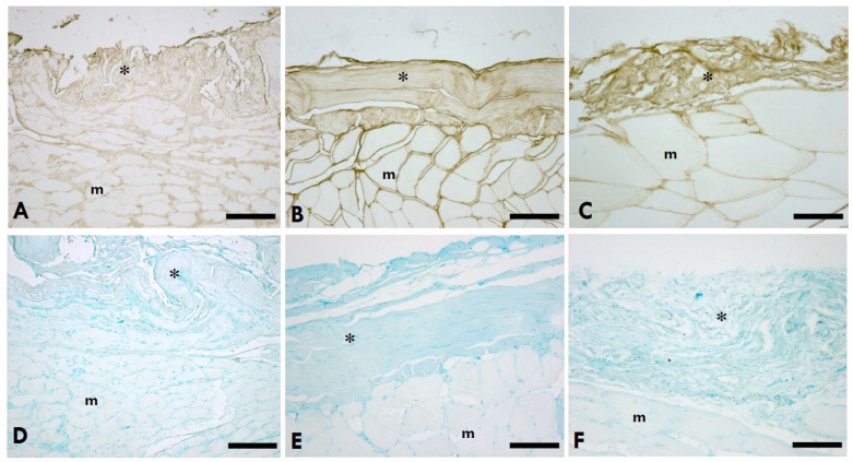
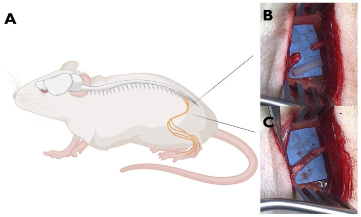
Peripheral nerve injury (PNI) is a complex clinical challenge resulting in functional disability. Neurological recovery does not always ensure functional recovery, as extracellular matrix (ECM) alterations affect muscle function. This study evaluates hyaluronan (HA) and collagen concentration in the gastrocnemius muscle and thoracolumbar fascia (TLF) in unilateral lower limb PNI rats to explore systemic ECM alterations following PNI and their impacts on functional recovery. Eighteen 8-week-old male Sprague-Dawley rats were divided into experimental (n = 12 left sciatic nerve injury) and control (n = 6) groups. After six weeks, motor function was evaluated. Muscle and TLF samples were analysed for HA and collagen distribution and concentrations. SFI and gait analysis confirmed a functional deficit in PNI rats 6 weeks after surgery. HA concentration in both sides of the muscles decreased by approximately one-third; both sides showed significantly higher collagen concentration than healthy rats (12.74 ± 4.83 µg/g), with the left (32.92 ± 11.34 µg/g) significantly higher than the right (20.15 ± 7.03 µg/g). PNI rats also showed significantly lower HA (left: 66.95 ± 20.08 µg/g; right: 112.66 ± 30.53 µg/g) and higher collagen (left: 115.89 ± 28.18 µg/g; right: 90.43 ± 20.83 µg/g) concentrations in both TLF samples compared to healthy rats (HA: 167.18 ± 31.13 µg/g; collagen: 47.51 ± 7.82 µg/g), with the left TLF more affected. Unilateral lower limb PNI induced HA reduction and collagen accumulation in both the lower limb muscles and the TLF, potentially exacerbating motor function impairment and increasing the risk of low back dysfunctions.

Keywords: collagen; extracellular matrix; hyaluronan; intramuscular connective tissue; peripheral nerve injury; thoracolumbar fascia.


Conflict of interest statement

The authors declare no conflicts of interest.

Figures

Figure 1
Figure 1
HA distribution in IMCT of the gastrocnemius muscle. (AC): Anti-HABP (hyaluronic acid binding protein); (DF): 0.05% Alcian Blue in MgCl2 0.05 M; (A,D) = left (sciatic nerve injury) side; (B,E) = right (contralateral) side; (C,F) = control (healthy rats); * = IMCT; m = muscle. Scale bars: 150 µm.
Figure 2
Figure 2
HA distribution in the TLF. (AC): Anti-HABP (hyaluronic acid binding protein); (DF): 0.05% Alcian Blue in MgCl2 0.05 M; (A,D) = left (sciatic nerve injury) side; (B,E) = right (contralateral) side; (C,F) = control (healthy rats); * = TLF; a = adipocytes. Scale bars: 150 µm.
Figure 3
Figure 3
Picrosirius Red staining of the gastrocnemius muscle. (A,D) = Left (sciatic nerve injury) side; (B,E) = right (contralateral) side; (C,F) = control—healthy rats. * = IMCT; m = muscle; e = endomysium; p = perimysium. Scale bars: (AC): 150 µm; (DF): 50 µm. Collagen is in red, the muscle fibres of the gastrocnemius muscle in yellow.
Figure 4
Figure 4
Picrosirius Red staining of the healthy TLF. (A): Scale bars 150 µm; (B): scale bars 50 µm; * = thoracolumbar fascia; a = adipocytes.
Figure 5
Figure 5
(A): The HA concentration in the gastrocnemius muscle on the left (sciatica nerve injury) side and right (contralateral) side of the left sciatic nerve injury rats and of the healthy rats (* p < 0.05). (B): The HA concentration in the TLF of the left (sciatica nerve injury) side and right (contralateral) side of the left sciatica nerve injury rats and of the healthy rats (** p < 0.01, *** p < 0.001). ANOVA with the Tukey post hoc test is applied to compare the difference between left and right sides of injured rats. Independent t-test is applied to compare the results of both sides of the injured rats and control separately.
Figure 6
Figure 6
(A): The collagen concentration in the gastrocnemius muscle of the left (sciatic nerve injury) side and the right (contralateral) side of the left sciatic nerve injury rats and of the healthy rats (* p < 0.05, *** p < 0.01). (B): The HA concentration in the TLF of the left (sciatic nerve injury) side and the right (contralateral) side of the left sciatica nerve injury rats and of the healthy rats (* p < 0.05, ** p < 0.01, *** p < 0.001). ANOVA with the Tukey post hoc test (for collagen concentration in the muscle) and Games–Howell post hoc test (for collagen concentration in the TLF) are applied to compare the difference between left and right sides of injured rats. Independent t-test is applied to compare the results of both sides of the injured rats and control separately.
Figure 7
Figure 7
The area percentage of collagen content in the gastrocnemius muscle of the left (sciatica nerve injury) side (n = 4) and the right (contralateral) side (n = 4) of the left sciatic nerve injury rats and of the healthy rats in the control group (n = 4). Kruskal–Wallis test with post hoc Mann–Whitney test is applied to compare the difference between left and right sides of injured rats and control separately (* p < 0.05).
Figure 8
Figure 8
Animal model of PNI. (A) Left sciatic nerve of the rat (created with BioRender.com) (B) Nerve exposure and gap (5 mm) creation. (C). Nerve repaired by interposition of an OxPVA-based nerve graft (10 mm) between the proximal and the distal stumps.
Figure 9
Figure 9
Experimental design.

Similar articles

References

    1. Aberg M., Ljungberg C., Edin E., Millqvist H., Nordh E., Theorin A., Terenghi G., Wiberg M. Clinical evaluation of a resorbable wrap-around implant as an alternative to nerve repair: A prospective, assessor-blinded, randomised clinical study of sensory, motor and functional recovery after peripheral nerve repair. J. Plast. Reconstr. Aesthet. Surg. 2009;62:1503–1509. doi: 10.1016/j.bjps.2008.06.041. - DOI - PubMed
    1. Menorca R.M., Fussell T.S., Elfar J.C. Nerve physiology: Mechanisms of injury and recovery. Hand Clin. 2013;29:317–330. doi: 10.1016/j.hcl.2013.04.002. - DOI - PMC - PubMed
    1. Lopes B., Sousa P., Alvites R., Branquinho M., Sousa A.C., Mendonça C., Atayde L.M., Luís A.L., Varejão A.S.P., Maurício A.C. Peripheral Nerve Injury Treatments and Advances: One Health Perspective. Int. J. Mol. Sci. 2022;23:918. doi: 10.3390/ijms23020918. - DOI - PMC - PubMed
    1. Carlson B.M. The Biology of Long-Term Denervated Skeletal Muscle. Eur. J. Transl. Myol. 2014;24:3293. doi: 10.4081/bam.2014.1.5. - DOI - PMC - PubMed
    1. Purslow P.P. The Structure and Role of Intramuscular Connective Tissue in Muscle Function. Front. Physiol. 2020;11:495. doi: 10.3389/fphys.2020.00495. - DOI - PMC - PubMed

Publication types

LinkOut - more resources