. 2024 Aug 17;25(16):8954.
doi: 10.3390/ijms25168954.

Protein Charge Neutralization Is the Proximate Driver Dynamically Tuning Reflectin Assembly

Affiliations

Protein Charge Neutralization Is the Proximate Driver Dynamically Tuning Reflectin Assembly

Robert Levenson et al. Int J Mol Sci. .

Abstract

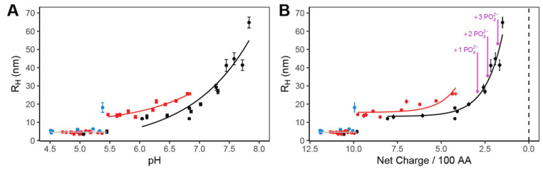
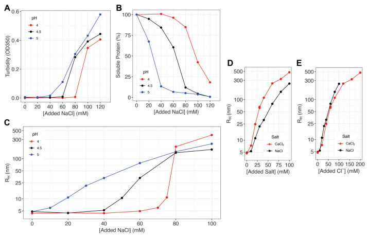
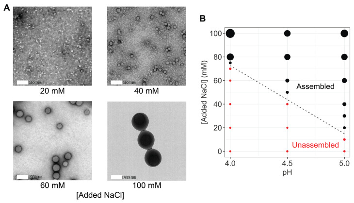
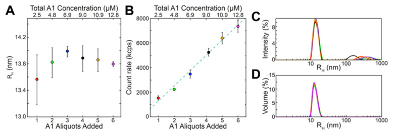
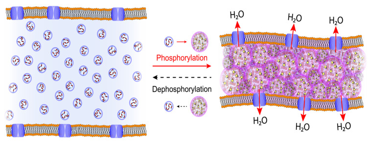
Reflectin is a cationic, block copolymeric protein that mediates the dynamic fine-tuning of color and brightness of light reflected from nanostructured Bragg reflectors in iridocyte skin cells of squids. In vivo, the neuronally activated phosphorylation of reflectin triggers its assembly, driving osmotic dehydration of the membrane-bounded Bragg lamellae containing the protein to simultaneously shrink the lamellar thickness and spacing while increasing their refractive index contrast, thus tuning the wavelength and increasing the brightness of reflectance. In vitro, we show that the reduction in repulsive net charge of the purified, recombinant reflectin-either (for the first time) by generalized anionic screening with salt or by pH titration-drives a finely tuned, precisely calibrated increase in the size of the resulting multimeric assemblies. The calculated effects of phosphorylation in vivo are consistent with these effects observed in vitro. The precise proportionality between the assembly size and charge neutralization is enabled by the demonstrated rapid dynamic arrest of multimer growth by a continual, equilibrium tuning of the balance between the protein's Coulombic repulsion and short-range interactive forces. The resulting stability of reflectin assemblies with time ensures a reciprocally precise control of the particle number concentration, encoding a precise calibration between the extent of neuronal signaling, osmotic pressure, and the resulting optical changes. The charge regulation of reflectin assembly precisely fine-tunes a colligative property-based nanostructured biological machine. A physical mechanism is proposed.

Keywords: biomaterials; intrinsically disordered proteins; protein assembly; reflectins.


Conflict of interest statement

The authors declare no conflicts of interest. Funders had no role in the design of the study; in the collection, analyses, or interpretation of data; in the writing of the manuscript; or in the decision to publish the results.

Figures

Figure 1
Figure 1
(A) Assembly sizes, measured by DLS, of D. opalescens reflectin A1 upon dilution into 25 mM buffers (blue = sodium acetate; red = MES; black = MOPS). A1 was dialyzed into 25 mM sodium acetate, pH 4.5, before pH neutralization to labeled pH conditions. Each data point corresponds to the mean of 10+ min of continuous DLS measurements of a freshly pH-neutralized sample. Error bars show the standard deviation of measurements. (B) Data from panel A, plotted as a function of calculated protein linear net charge density using the Henderson–Hasselbalch equation and reflectin sequence, as calculated previously [8,11]. Purple arrows indicate the calculated net charge density of reflectin A1 at pH 7 under varying degrees of phosphorylation.
Figure 2
Figure 2
(A) Turbidity (OD350) of reflectin A1 solutions in 25 mM sodium acetate as a function of pH and NaCl concentration. (B) The percentage of soluble protein remaining in the supernatant after the centrifugation of samples in A is calculated relative to the amount pre-centrifugation. (C) Sizes of majority populations of assembled reflectin assemblies as measured by DLS. Each data point is an average of 40 min continuous DLS measurements (see Methods). (D,E) Comparison of NaCl and CaCl2-induced assembly at pH 5.0 as a function of (D) salt concentration and (E) Cl-anion concentration. All experiments used 10 µM reflectin A1 filtered in 25 mM sodium acetate buffer pre-titrated to designated pH. The results shown are typical results from duplicate experiments.
Figure 3
Figure 3
(A) Transmission electron microscopy (TEM) images of negatively stained A1 in 25 mM acetate, pH 5.0, assembled in the presence of labeled NaCl concentrations. The 20 mM and 40 mM NaCl panels used 1 µM A1, while 60 mM 100 mM panels used 10 µM A1. (B) Reflectin A1 assembly as a function of pH and NaCl concentration. Point sizes are scaled to particle size as measured by DLS. For this panel, all particles with RH > 7.5 nm are considered assembled.
Figure 4
Figure 4
Reflectin assemblies are dynamically arrested and stable, as seen in these results of incremental additions of reflectin A1 monomers to the same 5 mM MOPS, pH 7.5 (neutralizing, assembly-driving) solution. (A) Reflectin assembly sizes (measured as the predominant DLS volume distributions; cf. Methods) as a function of monomer aliquots added. (B) Total scattering count rate as a function of monomer aliquots added (bottom x axis) and cumulative concentration (top x axis). Each point in A–B is the average of 3 replicate experiments, in each of which every aliquot addition was analyzed by 3 individual DLS measurements; error bars signify ± one S.D. between averages of replicates. Samples showed no significant variation over time following each aliquot addition, which is consistent with previous work [8,11]. Representative intensity (C) and volume (D) distributions observed after addition of the 6th aliquot of monomer with results after each aliquot shown in a different color.
Figure 5
Figure 5
Charge neutralization by phosphorylation (stars) triggers a precisely limited assembly of the cationic, block-copolymeric protein, reflectin, driving a calibrated osmotic tuning of color reflected from Bragg lamellae in cells of squid skin. Effective neutralization by anionic screening provides a surrogate for phosphorylation, driving reflectin’s proportional assembly in vitro.

Similar articles

Cited by

References

    1. Hanlon R. Cephalopod Dynamic Camouflage. Curr. Biol. 2007;17:R400–R404. doi: 10.1016/j.cub.2007.03.034. - DOI - PubMed
    1. Izumi M., Sweeney A.M., DeMartini D., Weaver J.C., Powers M.L., Tao A., Silvas T.V., Kramer R.M., Crookes-Goodson W.J., Mäthger L.M., et al. Changes in Reflectin Protein Phosphorylation Are Associated with Dynamic Iridescence in Squid. J. R. Soc. Interface. 2010;7:549–560. doi: 10.1098/rsif.2009.0299. - DOI - PMC - PubMed
    1. Tao A.R., DeMartini D.G., Izumi M., Sweeney A.M., Holt A.L., Morse D.E. The Role of Protein Assembly in Dynamically Tunable Bio-Optical Tissues. Biomaterials. 2010;31:793–801. doi: 10.1016/j.biomaterials.2009.10.038. - DOI - PubMed
    1. DeMartini D.G., Krogstad D.V., Morse D.E. Membrane Invaginations Facilitate Reversible Water Flux Driving Tunable Iridescence in a Dynamic Biophotonic System. Proc. Natl. Acad. Sci. USA. 2013;110:2552–2556. doi: 10.1073/pnas.1217260110. - DOI - PMC - PubMed
    1. Ghoshal A., DeMartini D.G., Eck E., Morse D.E. Optical Parameters of the Tunable Bragg Reflectors in Squid. J. R. Soc. Interface. 2013;10:20130386. doi: 10.1098/rsif.2013.0386. - DOI - PMC - PubMed