. 2024 Aug 17;25(16):8959.
doi: 10.3390/ijms25168959.

Functional and Structural Changes in Diaphragm Neuromuscular Junctions in Early Aging

Affiliations

Functional and Structural Changes in Diaphragm Neuromuscular Junctions in Early Aging

Andrei N Tsentsevitsky et al. Int J Mol Sci. .

Abstract

Age-related impairment of the diaphragm causes respiratory complications. Neuromuscular junction (NMJ) dysfunction can be one of the triggering events in diaphragm weaknesses in old age. Prominent structural and functional alterations in diaphragm NMJs were described in elderly rodents, but NMJ changes in middle age remain unclear. Here, we compared diaphragm muscles from young adult (3 months) and middle-aged (12 months) BALB/c mice. Microelectrode recordings, immunofluorescent staining, electron microscopy, myography, and whole-body plethysmography were used. We revealed presynaptic (i) and postsynaptic (ii) changes. The former (i) included an increase in both action potential propagation velocity and neurotransmitter release evoked by low-, moderate-, and high-frequency activity but a decrease in immunoexpression of synapsin 1 and synaptic vesicle clustering. The latter (ii) consisted of a decrease in currents via nicotinic acetylcholine receptors and the area of their distribution. These NMJ changes correlated with increased contractile responses to moderate- to high-frequency nerve activation. Additionally, we found alterations in the pattern of respiration (an increase in peak inspiratory flow and a tendency of elevation of the tidal volume), which imply increased diaphragm activity in middle-aged mice. We conclude that enhancement of neuromuscular communication (due to presynaptic mechanism) accompanied by improved contractile responses occurs in the diaphragm in early aging.

Keywords: acetylcholine; aging; contraction; diaphragm; end-plate; neuromuscular junction; neurotransmitter release; respiration; synapsin; synaptic vesicle.


Conflict of interest statement

The authors declare no conflict of interest.

Figures

Figure 1
Figure 1
Measurements of latency and synaptic delay. (A) Typical extracellular recording, including artifact, AP, and EPP, is shown. Intervals corresponding to latencies (Lat. and Lat.S) and synaptic delay (Syn. Del.) are denoted as a, b, and c, respectively. Insert, the representative recordings from NMJs of 3- and 12-month-old mice (3 mo and 12 mo). (B) Quantification of latencies and synaptic delay. Data are represented as mean ± standard deviation (SD). n = 64 and 33 for 3 and 12 months. * p < 0.05, ** p < 0.01 *** p < 0.001 by Mann–Whitney U-test between groups.
Figure 2
Figure 2
Spontaneous and evoked ACh release. (A) Typical traces of MEPPs and EPPs recorded extracellularly in 3- and 12-month-old mice (3 mo and 12 mo). Three traces are overlaid in each example. Quantification of MEPP frequency (B), quantal content (m) of EPPs (C), and ratio (Rt/rt) of MEPP and EPP rise times (D). Data are represented as mean ± SD. n = 67–68 and 37–38 for 3 and 12 months. *** p < 0.001 by Mann–Whitney U-test between groups; ns—non-significant.
Figure 3
Figure 3
Parameters of miniature postsynaptic responses. (A) Amplitude, (B) rise time, and (C) decay time of MEPPs. n = 68 and 38 for 3 and 12 months. Data are represented as mean ± SD. *** p < 0.001 by Mann–Whitney U-test between groups; ns—non-significant.
Figure 4
Figure 4
Evoked ACh release at different modes of phrenic nerve activation. (A) Changes in quantal content during 3 min intermittent stimulation at 20 Hz. Averaged curves are shown. (B) Cumulative quantal content indicating a number of quanta released for 3 min of intermittent stimulation at 20 Hz. (C) Dynamic neurotransmitter release upon shot stimulus trains at 10, 20, 50, and 70 Hz. Insert, graphs show the number of quanta released during these stimulus trains. (D) Estimation of readily releasable pool (RRP) size by plotting quantal content (m; the y-axis) over the cumulative quantal content (x-axis) and extrapolating the initial linear decay phase on x-axis. Data are represented as mean ± SD. n = 11 and 7 for 3 and 12 months. * p < 0.05 and *** p < 0.001 by Mann–Whitney U-test between groups.
Figure 5
Figure 5
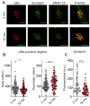
Labeling of postsynaptic and presynaptic components of NMJs. (A) The representative fluorescence images of NMJs, where postsynaptic nAChRs and presynaptic proteins (synapsin 1 and anti-SNAP-25) were labeled with αBtx and specific antibodies, respectively. Scale bars—25 µm. (B) Estimation of αBtx-positive area and perimeter. (C) Quantification of synapsin 1 immunofluorescence. Data are represented as mean ± SD; 143 NMJs from n = 7 (3 months) and 102 NMJ from n = 5 (12 months) were analyzed. * p < 0.05 and *** p < 0.001 by Mann–Whitney U-test between groups.
Figure 6
Figure 6
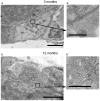
Electron microscopy of NMJs. (a,b) Representative TEM images of NMJs from 3-month-old and 12-month-old mice. (a’,b’) Regions containing active zone, synaptic vesicle cluster, postsynaptic infoldings, and synaptic cleft at a higher magnification. Scale bars—0.5 µm (a’,b’) and 2 µm (a,b).
Figure 7
Figure 7
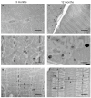
Ultrastructure of DIAm myofibrils of 3-month-old and 12-month-old mice. TEM images: (a,b) general view of muscle fiber bundles; (c,d) cross-section of myofibrils; (e,f) longitudinal section of myofibrils, sarcomere structure. Scale bars—5 µm (a,b) and 1 µm other microphotographs. Abbreviations: A—A-band, I—I-band, lb—lipid body, mt—mitochondria, n—nucleus, S—sarcomere, Z—Z-line.
Figure 8
Figure 8
Muscle contractions evoked by nerve activation. (A) Representative traces of contractile responses to low- (0.1 Hz), moderate- (20 Hz), and high- (70 Hz) frequency stimulation in 3- and 12-month-old mice. (B) Quantification of maximal contraction force elicited by phrenic nerve stimulation at 0.1, 10, 20, 50, and 70 Hz. Data are represented as mean ± SD. n = 11 and 8 for 3 and 12 months. ns—non-significant and ** p < 0.01 by Mann–Whitney U-test between groups.
Figure 9
Figure 9
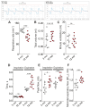
Whole-body plethysmography of 3- and 12-month-old mice. Top, the representative recordings. Quantification of respiratory rate (A), tidal volume (B), and minute ventilation (C) are shown. (DF) Graphs show the kinetic parameters of breathing: inspiratory and expiratory time (D), peak inspiratory and expiratory flows (E), and mid-tidal expiratory flow or EF50 (F). Data are represented as mean ± SD. n = 10 and 11 for 3 and 12 months. * p < 0.05 and *** p < 0.001 by Mann–Whitney U-test between groups; ns, non-significant.

References

    1. Elliott J.E., Greising S.M., Mantilla C.B., Sieck G.C. Functional impact of sarcopenia in respiratory muscles. Respir. Physiol. Neurobiol. 2016;226:137–146. doi: 10.1016/j.resp.2015.10.001. - DOI - PMC - PubMed
    1. Bordoni B., Morabito B., Simonelli M. Ageing of the Diaphragm Muscle. Cureus. 2020;12:e6645. doi: 10.7759/cureus.6645. - DOI - PMC - PubMed
    1. Saldarriaga C.A., Alatout M.H., Khurram O.U., Gransee H.M., Sieck G.C., Mantilla C.B. Chloroquine impairs maximal transdiaphragmatic pressure generation in old mice. J. Appl. Physiol. 2023;135:1126–1134. doi: 10.1152/japplphysiol.00365.2023. - DOI - PMC - PubMed
    1. Siniscalchi C., Nouvenne A., Cerundolo N., Meschi T., Ticinesi A. On Behalf of The Parma Post-Graduate Specialization School in Emergency-Urgency Medicine Interest Group on Thoracic, U. Diaphragm Ultrasound in Different Clinical Scenarios: A Review with a Focus on Older Patients. Geriatrics. 2024;9:70. doi: 10.3390/geriatrics9030070. - DOI - PMC - PubMed
    1. Greising S.M., Mantilla C.B., Gorman B.A., Ermilov L.G., Sieck G.C. Diaphragm muscle sarcopenia in aging mice. Exp. Gerontol. 2013;48:881–887. doi: 10.1016/j.exger.2013.06.001. - DOI - PMC - PubMed

LinkOut - more resources