. 2024 Aug 17;25(16):8968.
doi: 10.3390/ijms25168968.

Generic Reporter Sets for Colorimetric Multiplex dPCR Demonstrated with 6-Plex SNP Quantification Panels

Affiliations

Generic Reporter Sets for Colorimetric Multiplex dPCR Demonstrated with 6-Plex SNP Quantification Panels

Maximilian Neugebauer et al. Int J Mol Sci. .

Abstract

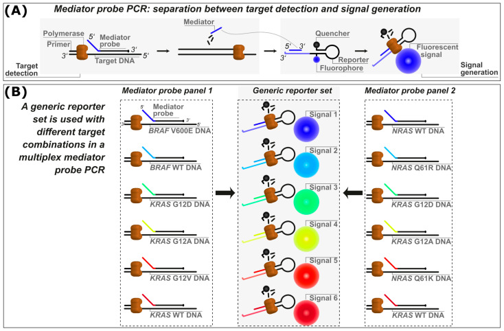
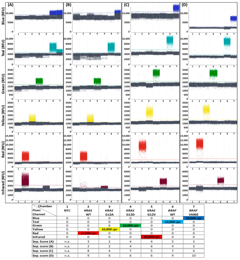
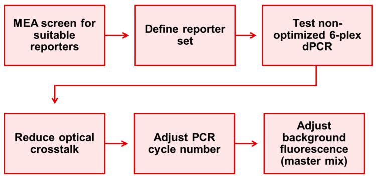
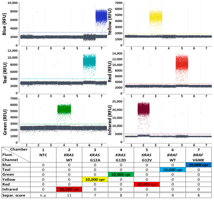
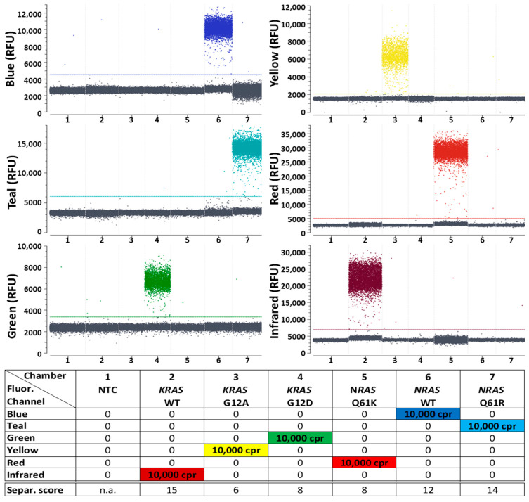
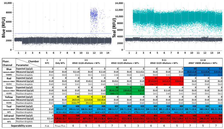
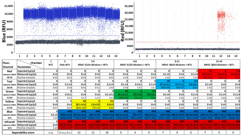
Digital PCR (dPCR) is a powerful method for highly sensitive and precise quantification of nucleic acids. However, designing and optimizing new multiplex dPCR assays using target sequence specific probes remains cumbersome, since fluorescent signals must be optimized for every new target panel. As a solution, we established a generic fluorogenic 6-plex reporter set, based on mediator probe technology, that decouples target detection from signal generation. This generic reporter set is compatible with different target panels and thus provides already optimized fluorescence signals from the start of new assay development. Generic reporters showed high population separability in a colorimetric 6-plex mediator probe dPCR, due to their tailored fluorophore and quencher selection. These reporters were further tested using different KRAS, NRAS and BRAF single-nucleotide polymorphisms (SNP), which are frequent point mutation targets in liquid biopsy. We specifically quantified SNP targets in our multiplex approach down to 0.4 copies per microliter (cp/µL) reaction mix, equaling 10 copies per reaction, on a wild-type background of 400 cp/µL for each, equaling 0.1% variant allele frequencies. We also demonstrated the design of an alternative generic reporter set from scratch in order to give detailed step-by-step guidance on how to systematically establish and optimize novel generic reporter sets. Those generic reporter sets can be customized for various digital PCR platforms or target panels with different degrees of multiplexing.

Keywords: BRAF; KRAS; NRAS; assay development; color compensation; digital PCR (dPCR); fluorophore combination; generic reporter set; mediator extension assay (MEA); mediator probe PCR (MP PCR); oncogenic mutations.


Conflict of interest statement

The authors declare no conflicts of interest. The funders had no role in the design of the study; in the collection, analyses, or interpretation of data; in the writing of the manuscript; or in the decision to publish the results.

Figures

Figure 1
Figure 1
(A) Schematic overview of the mediator probe PCR (MP PCR) principle, showing the separation between the target detection and fluorescence signal generation steps. (B) Schematic overview of the generic reporter set principle. A generic reporter set with highly distinguishable fluorophore signals is designed and optimized. Each of these reporters needs to be tagged with a specific mediator binding site sequence to allow binding of a specific mediator. Mediator probes (MPs) with target binding sites and mediator sequences that are reverse complementary to the mediator binding sites of the reporter as well as appropriate primer pairs are designed (mediator probe panel 1). The mediators are released after being cleaved from their MP by polymerase and bind the corresponding mediator binding site for signal generation. Several mediator probe panels (e.g., mediator probe panel 2) with the same mediator sequences can be designed for combination with the generic reporter set. WT: wild-type.
Figure 6
Figure 6
Improving discriminability between positive and negative signal populations in dPCR reactions step-by-step. During all steps of generic reporter set optimization, the target panel for detection of KRAS and BRAF mutations and their corresponding WT controls was used. 1 D-plots of all 6 fluorescence channels are shown. Reaction chamber details from left to right: (1) NTC; (2–7) 10,000 cpr of one kind of target sequence each. A more detailed version of this figure, with samples containing all six targets at once, is shown in Figure S5. Separability scores between positive and negative droplet populations are indicated at the bottom of the legend. Fluorophores for this second generic reporter set were selected according to the MEA data shown in Figure S3. Details of the used reporters in each detection channel including sequences, fluorophores and quenchers can be seen in Table S2. (A) Initial non-optimized test without color compensation. (B) Initial non-optimized test after setting a suitable color compensation, with which each target is detected in only one fluorescence channel. This color compensation matrix was henceforth used in every subsequent test to optimize the second generic reporter set. (C) Improved separability after increasing the PCR cycle number from 45 to 60. (D) Further improved separability after reduction of background fluorescence in the “Blue” and “Teal” channels by changing the naica® master mix version from “naica® multiplex PCR MIX” to “naica® PCR MIX”. Although the second generic reporter set shown here is an optimized set, ready to be used in combination with other target panels, it utilizes different reporters from the first 6-plex generic reporter set that was characterized in the KRAS, BRAF and NRAS quantification studies shown in Section 2.1.
Figure 7
Figure 7
6-plex dPCR assay optimization workflow for definition of a generic reporter set as performed in this work and recommended for the establishment of new generic reporter sets.
Figure 2
Figure 2
Differentiation between positive and negative signal populations in Digital PCR (dPCR) reactions using the first 6-plex generic reporter set in combination with a target panel for detection of KRAS and BRAF mutations and their corresponding WT controls. One-dimensional plots of all 6 fluorescence channels (labeled “Blue”, Teal”, “Green”, “Yellow”, “Red” and “Infrared”) of the naica® Prism6 are shown. Details of the reporters used in each detection channel, including sequences, fluorophores and quenchers, can be seen in Table S1. All detectable droplets formed in seven separate reaction chambers are shown on the x-axis, and the relative fluorescence units (RFU) of each droplet are shown on the y-axis. Reaction chamber details from left to right: (1) no template control (NTC) without target molecule presence; (2–7) 10,000 copies per reaction (cpr) of one kind of target sequence each. A more detailed version of this figure, with samples containing all six targets at once, is shown in Figure S1. Separability scores between positive and negative droplet populations, as calculated by the Crystal Miner software, are indicated at the bottom of the legend. n.a.: not applicable.
Figure 3
Figure 3
Differentiation between positive and negative signal populations in dPCR reactions using the first 6-plex generic reporter set in combination with a target panel for detection of KRAS and NRAS mutations and their corresponding WT controls. One-dimensional plots of all 6 fluorescence channels are shown. Details of the reporters used in each detection channel, including sequences, fluorophores and quenchers, can be seen in Table S1. Reaction chamber details from left to right: (1) NTC; (2–7) 10,000 cpr of one kind of target sequence each. A more detailed version of this figure, with samples containing all six targets at once, is shown in Figure S2. Separability scores between positive and negative droplet populations are indicated at the bottom of the legend.
Figure 4
Figure 4
Quantitative dPCR results of the first 6-plex generic reporter set in combination with a target panel for detection of KRAS and BRAF mutations and their corresponding WT controls. A background of 10,000 cpr of both WT targets is present in all reaction chambers aside from the NTC (reaction chamber 1). Single mutation targets were added in three different concentrations (1000 cpr, 100 cpr and 10 cpr). Above: One-dimensional plots of fluorescence channels detecting either BRAF WT in “Teal” or BRAF V600E in “Blue”. One-dimensional plots of all other fluorescence channels detecting the other targets are depicted in Figure S6. Below: Quantitative data of three replicates with reactions freshly prepared for each experiment. Expected concentrations, mean values and standard deviations of measured concentrations in copies per microliter (cp/μL) as well as mean values and standard deviations of the number of positive droplets in all three experiments are shown for each reaction chamber in each detection channel. Cells are color-coded wherever a positive result was expected for the specific chamber and channel. Separability scores between positive and negative droplet populations in the depicted reactions are indicated at the bottom.
Figure 5
Figure 5
Quantitative PCR results of the first 6-plex generic reporter set in combination with a target panel for detection of KRAS and NRAS mutations and their corresponding WT controls. A background of 10,000 cpr of both WT targets is present in all reaction chambers aside from the NTC (reaction chamber 1). Single mutation targets were added in three different concentrations (1000 cpr, 100 cpr and 10 cpr). Above: One-dimensional plots of fluorescence channels detecting either NRAS WT in “Blue” or NRAS Q61K in “Red”. One-dimensional plots of all other fluorescence channels detecting the other targets are depicted in Figure S7. Below: Quantitative data of three replicates with reactions freshly prepared for each experiment. Expected concentrations, mean values and standard deviations of measured concentrations in cp/µL as well as mean values and standard deviations of the number of positive droplets in all three experiments are shown for each reaction chamber in each detection channel. Cells are color-coded wherever a positive result was expected for the specific chamber and channel. Separability scores between positive and negative droplet populations in the depicted reactions are indicated at the bottom.

Similar articles

Cited by

References

    1. Olmedillas-López S., Olivera-Salazar R., García-Arranz M., García-Olmo D. Current and Emerging Applications of Droplet Digital PCR in Oncology: An Updated Review. Mol. Diagn. Ther. 2022;26:61–87. doi: 10.1007/s40291-021-00562-2. - DOI - PubMed
    1. Fang W., Liu X., Maiga M., Cao W., Mu Y., Yan Q., Zhu Q. Digital PCR for Single-Cell Analysis. Biosensors. 2024;14:64. doi: 10.3390/bios14020064. - DOI - PMC - PubMed
    1. Taylor S.C., Laperriere G., Germain H. Droplet Digital PCR versus qPCR for gene expression analysis with low abundant targets: From variable nonsense to publication quality data. Sci. Rep. 2017;7:2409. doi: 10.1038/s41598-017-02217-x. - DOI - PMC - PubMed
    1. Hou Y., Chen S., Zheng Y., Zheng X., Lin J.-M. Droplet-based digital PCR (ddPCR) and its applications. TrAC Trends Anal. Chem. 2023;158:116897. doi: 10.1016/j.trac.2022.116897. - DOI
    1. Whale A.S., Huggett J.F., Tzonev S. Fundamentals of multiplexing with digital PCR. Biomol. Detect. Quantif. 2016;10:15–23. doi: 10.1016/j.bdq.2016.05.002. - DOI - PMC - PubMed

MeSH terms

LinkOut - more resources