. 2024 Aug 19;25(16):8994.
doi: 10.3390/ijms25168994.

The Regulation of MicroRNA-21 by Interleukin-6 and Its Role in the Development of Fibrosis in Endometriotic Lesions

Affiliations

The Regulation of MicroRNA-21 by Interleukin-6 and Its Role in the Development of Fibrosis in Endometriotic Lesions

Maria Ariadna Ochoa Bernal et al. Int J Mol Sci. .

Abstract

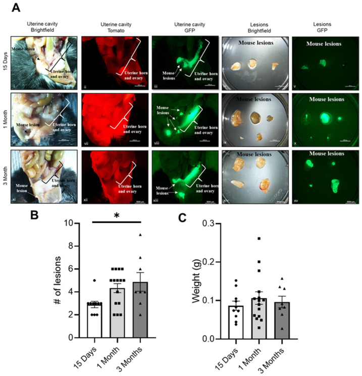
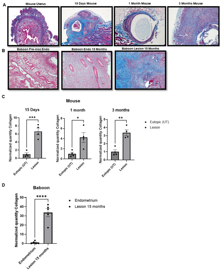
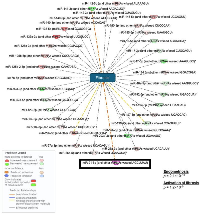
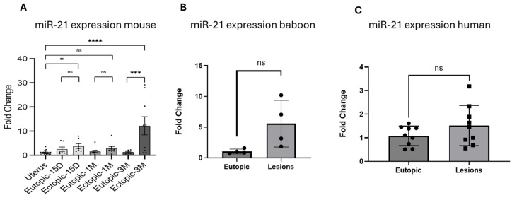
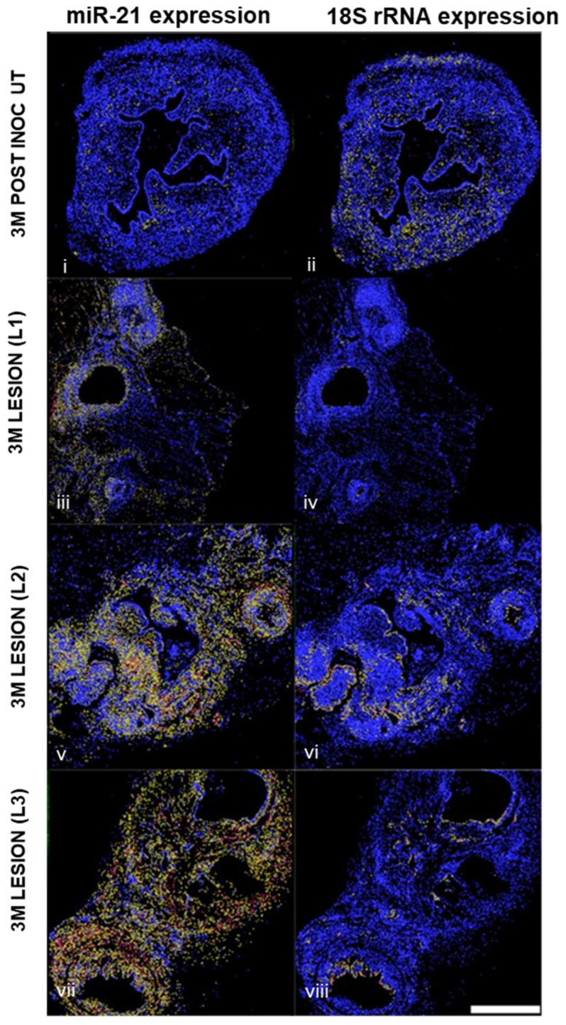
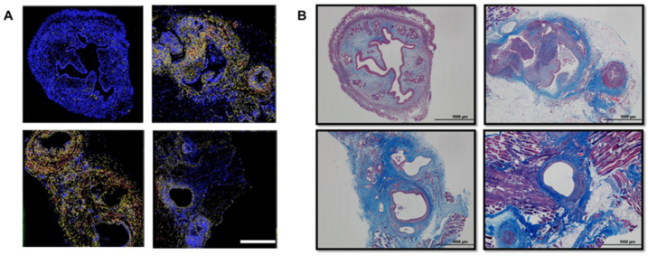
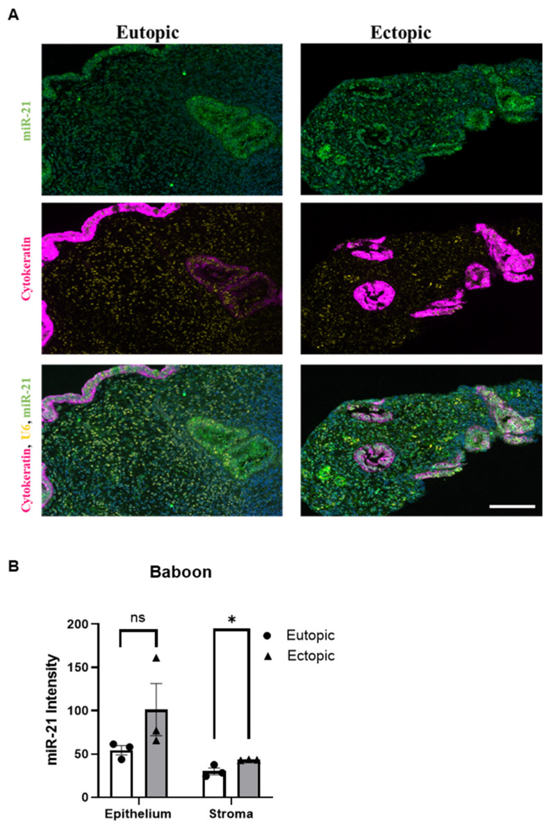
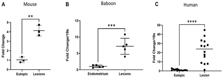
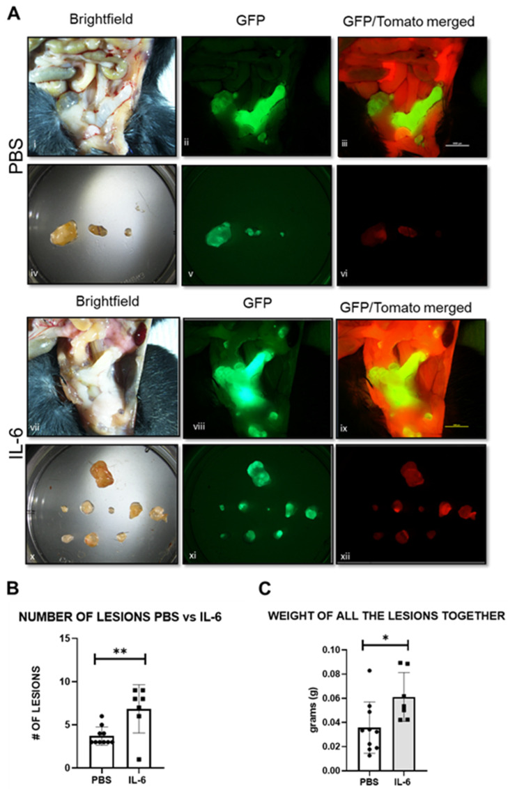
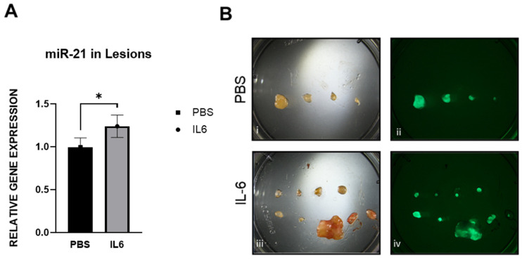
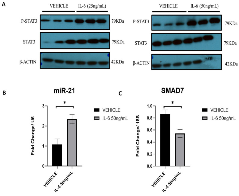
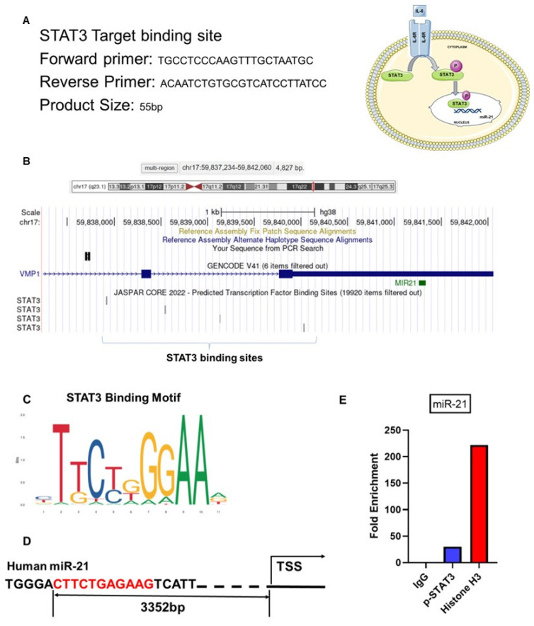
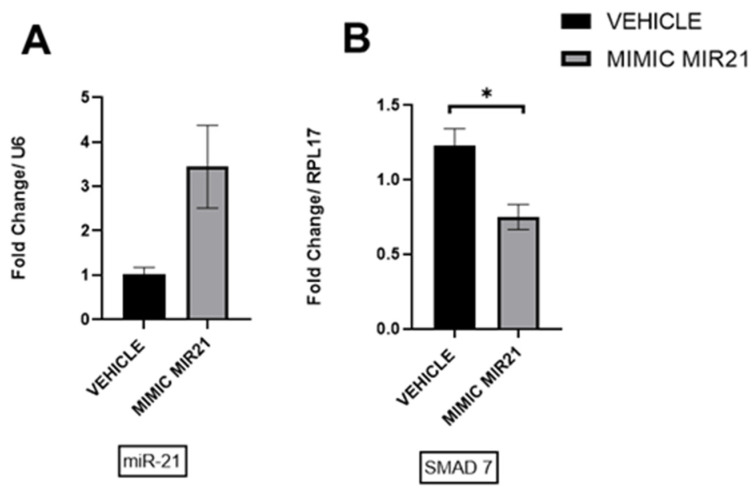
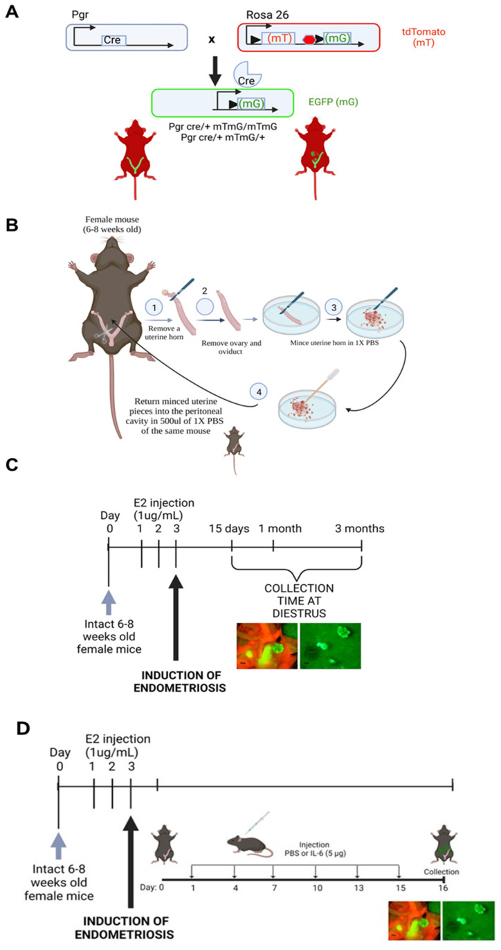
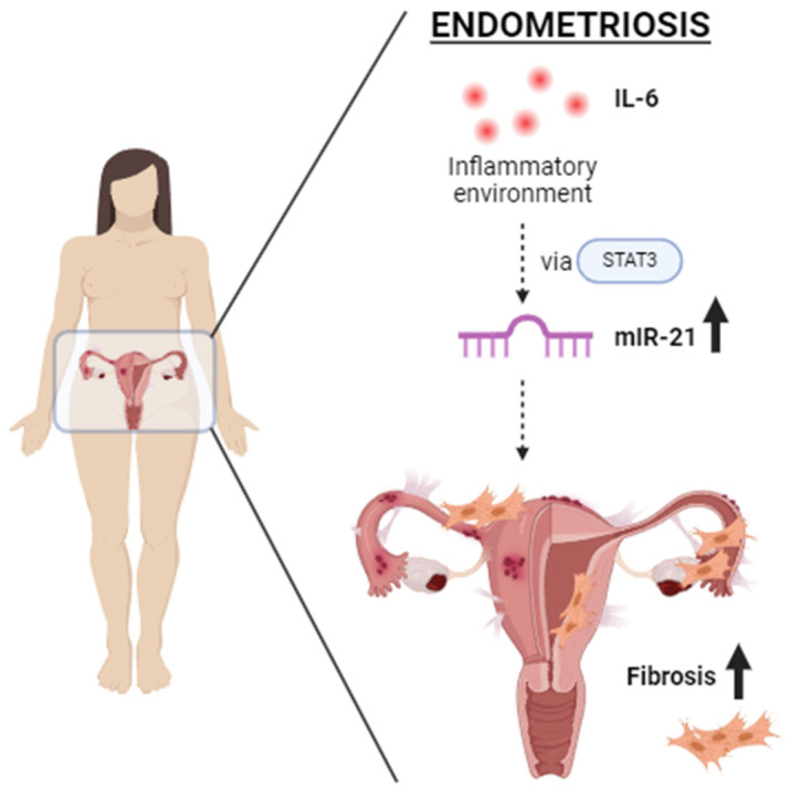
Endometriosis is one of the most common causes of chronic pelvic pain and infertility that affects 10% of women of reproductive age. It is currently defined as the presence of endometrial epithelial and stromal cells at ectopic sites; however, advances in endometriosis research have some authors believing that endometriosis should be re-defined as "a fibrotic condition in which endometrial stroma and epithelium can be identified". microRNAs (miRNAs) are regulatory molecules that potentially play a role in endometriotic lesion development. There is evidence that suggests that miRNAs, including microRNA-21 (miR-21), participate in fibrotic processes in different organs, including the heart, kidney, liver and lungs. The objective of this study was to understand the role of miR-21 and the mechanisms that can contribute to the development of fibrosis by determining how IL-6 regulates miR-21 expression and how this miRNA regulates the transforming growth factor beta (TGF-β) signaling pathway to promote fibrosis. We investigated the expression of miR-21 in the baboon and mouse model of endometriosis and its correlation with fibrosis. We demonstrated that inflammation and fibrosis are present at a very early stage of endometriosis and that the inflammatory environment in the peritoneal cavity, which includes interleukin 6 (IL-6), can regulate the expression of miR-21 in vitro and in vivo.

Keywords: IL-6; endometriosis; fibrosis; inflammation; microRNA-21.


Conflict of interest statement

The authors declare no conflicts of interest.

Figures

Figure 1
Figure 1
Progression of endometriosis using the Pgr cre/+ Rosa26 mT/mG mouse model: (A) Brightfield, Tomato red and GFP images of the uterine cavity of the mouse at 15 days, 1 month and 3 months after induction of endometriosis. Lesions collected from those animals are shown with brightfield and GFP. Scale bar: 5000 μm. (B) Comparison of the number of lesions. * p < 0.05 and (C) weight of the lesions over time. Mice at 15 days (n = 10), 1 month (n = 15) and 3 months (n = 8).
Figure 2
Figure 2
Masson’s trichrome staining: (A) Mouse uterus and 15 days (n = 4), 1 month (n = 4) and 3-month lesions (n = 4) Deposition of collagen (blue) around the endometriotic lesion. (B) Baboon pre-inoculation endometrium, endometrium after 15 months with endometriosis and 15-month endometriotic lesion (n = 5). Deposition of collagen (blue) around the endometriotic lesion. Scale bar: 100 μm. (C) Collagen quantification in the mouse model of endometriosis at 15 days, 1 month and 3 months. (D) Collagen quantification in the baboon model of endometriosis. * p < 0.05, ** p < 0.01, *** p < 0.001 and **** p < 0.0001.
Figure 3
Figure 3
Differentially expressed (DE) miRNAs associated with activation of fibrosis in endometriosis between eutopic and ectopic endometrium in 15-month baboon samples. Small RNA-seq analysis shows the most relevant miRNAs involved in the development of fibrosis, including miR-21. All baboon samples combined, n = 24. (*) Multiple DE microRNAs identified within the same familiy of microRNAs.
Figure 4
Figure 4
miR-21 expression in mouse, baboon and human lesions: (A) mir-21 fold change comparing uterine lesions and matched eutopic lesions in mice at 15 days (n = 7), 1 month (n = 10) and 3 months (n = 9) with endometriosis; (B) baboons at 15 months after endometriosis induction (n = 4), (C) women (n = 9) with endometriosis. ns (not significant), * p < 0.05, *** p < 0.001 and **** p < 0.0001.
Figure 5
Figure 5
In situ hybridization of miR-21 expression and 18SrRNA expression in mouse uterus and lesions (n = 3). Staining intensity was displayed in blue, yellow or red depending on their low to high intensity. Scale bar: 500 μm.
Figure 6
Figure 6
Correlation of miR-21 expression with fibrosis in mouse uterus and three months mouse lesions: (A) In situ hybridization of miR-21. miR-21 is present in the stromal cells within the endometriotic lesions; (B) Masson’s trichrome staining of the adjacent mouse sections of the uterus and 3-month lesions. Note the deposition of collagen (blue) within the endometriotic lesion. (n = 3) Scale bar: 1000 μm.
Figure 7
Figure 7
In situ hybridization of miR-21 in the baboon model: (A) The left panel shows the in situ hybridization of the eutopic endometrium of the baboon and the right panel shows an ectopic lesion. miR-21 is represented in green, cytokeratin in pink and U6 in yellow. (n = 3). Scale bar 300 px. (B) miR-21 quantification in eutopic and ectopic . ns (not significant), * p < 0.05.
Figure 8
Figure 8
Expression of CTGF in (A) mouse, (B) baboon and (C) women with endometriosis. A significant increase in CTGF expression in lesions was evident in mouse (n = 3), baboon (n = 5) and women with endometriosis (n = 14). ** p < 0.01, *** p < 0.001 and **** p < 0.0001.
Figure 9
Figure 9
Mouse lesions and uterus following PBS and IL-6 treatment: (A) Representative pictures in brightfield (i,iv,vii,x) and fluoresce GFP (ii,v,vii,xi) and GFP/Tomato merged (iii,vi,ix,xii). (B) Number of lesions in mice injected with PBS (n = 10) or IL-6 (n = 7). (C) Weight of all the lesions pooled together in animals injected with PBS or IL-6. * p < 0.05, ** p < 0.01.
Figure 10
Figure 10
Comparison of miR-21 expression between mice injected with PBS and IL-6: (A) Expression of miR-21 in PBS (n = 4) compared with IL-6 treated mice (n = 3) (B) Representative pictures of lesions from PBS and IL-6 mice in brightfield (i,iii) and under fluorescence GFP (ii,iv). * p <0.05.
Figure 11
Figure 11
Regulation of miR-21 expression via P-STAT3: (A) Western Blot at 24 h of p-STAT3 and STAT3 in ectopic stromal cells in the presence of recombinant IL-6 at 25 ng/mL and 50 ng/mL. (B) RT-qPCR analysis at 12 h of the miR-21 expression. (C) RT-qPCR analysis of the miR-21 target, SMAD7 expression (n = 3). * p < 0.05.
Figure 12
Figure 12
Predicted Transcription Factor binding sites for STAT3: (A) The primer used for the binding site. (B) The results from the transcription factor binding profile database JASPAR CORE 2022. The present study suggests that the expression of miR-21 is increased in ectopic stromal cells via p-STAT3 binding. (C,D) The predicted STAT3 binding site of human miR-21 promoter. (E) The binding efficiency of STAT3 on the human miR-21 promoter enhanced by IL-6 stimulation.
Figure 13
Figure 13
Overexpression of miR-21 in ectopic stromal cells: (A) Upregulation of miR-21 (B) was associated with the downregulation of Smad7 mRNA (n = 3). * p < 0.05.
Figure 14
Figure 14
Mouse model used for the induction of endometriosis: (A) Schematic diagram of the mT/mG mouse construct before and after Cre-mediated recombination. (B) Diagram illustrating the surgical procedure for the induction of endometriosis using the double-fluorescent Cre reporter mouse. (C) Schematic diagram of the induction of endometriosis in the mT/mG mouse model of endometriosis. Lesions for the analysis were collected at 15 days, 1 month and 3 months post-induction during diestrus. (D) Experimental design of the mouse model of endometriosis and the days of injection of IL-6 or PBS after endometriosis induction. Created with BioRender.com.
Figure 15
Figure 15
Proposed hypothesis: The inflammatory environment, particularly IL-6, in the peritoneal cavity of women with endometriosis can upregulate miR-21 via STAT3, leading to an increase in fibrosis in endometriotic lesions.

Similar articles

Cited by

References

    1. Giudice L.C., Kao L.C. Endometriosis. Lancet. 2004;364:1789–1799. doi: 10.1016/S0140-6736(04)17403-5. - DOI - PubMed
    1. Giudice L.C. Endometriosis. N. Engl. J. Med. 2010;362:2389–2398. doi: 10.1056/NEJMcp1000274. - DOI - PMC - PubMed
    1. Vigano P., Candiani M., Monno A., Giacomini E., Vercellini P., Somigliana E. Time to redefine endometriosis including its pro-fibrotic nature. Hum. Reprod. 2018;33:347–352. doi: 10.1093/humrep/dex354. - DOI - PubMed
    1. Bulun S.E. Endometriosis. N. Engl. J. Med. 2009;360:268–279. doi: 10.1056/NEJMra0804690. - DOI - PubMed
    1. Zondervan K.T., Becker C.M., Koga K., Missmer S.A., Taylor R.N., Vigano P. Endometriosis. Nat. Rev. Dis. Primers. 2018;4:9. doi: 10.1038/s41572-018-0008-5. - DOI - PubMed