Review
. 2024 Aug 20;25(16):9042.
doi: 10.3390/ijms25169042.

The Synergistic Effects of Polyol Pathway-Induced Oxidative and Osmotic Stress in the Aetiology of Diabetic Cataracts

Affiliations
Review

The Synergistic Effects of Polyol Pathway-Induced Oxidative and Osmotic Stress in the Aetiology of Diabetic Cataracts

Courtney A Thorne et al. Int J Mol Sci. .

Abstract

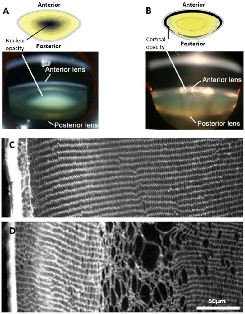
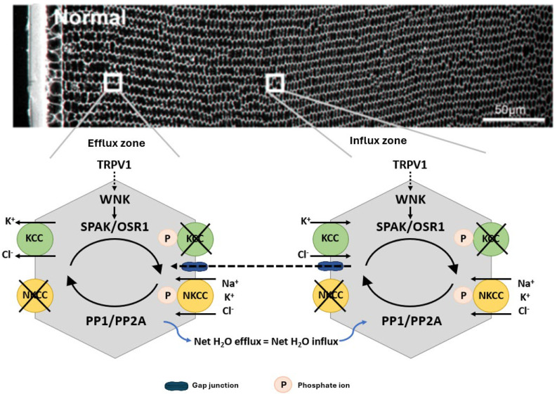
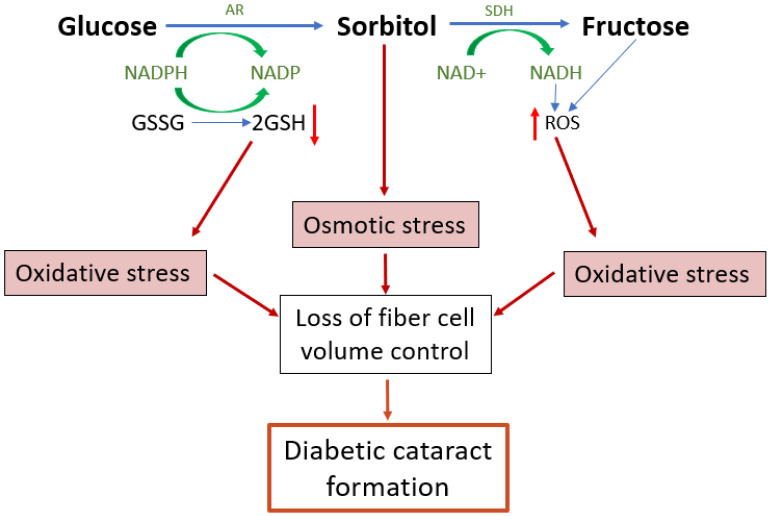
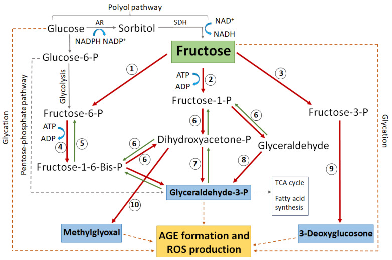
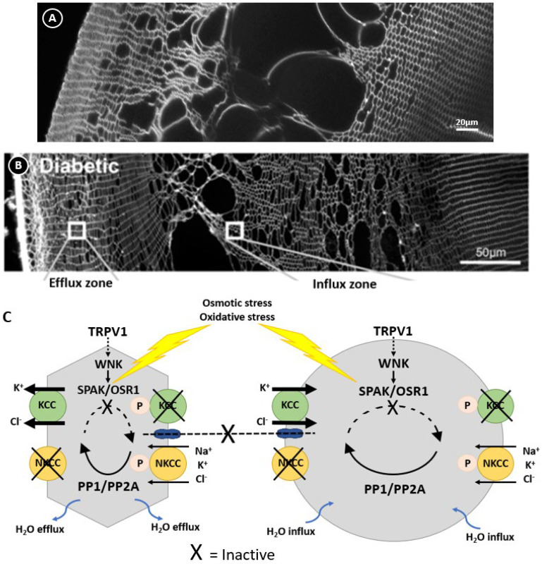
Cataracts are the world's leading cause of blindness, and diabetes is the second leading risk factor for cataracts after old age. Despite this, no preventative treatment exists for cataracts. The altered metabolism of excess glucose during hyperglycaemia is known to be the underlying cause of diabetic cataractogenesis, resulting in localised disruptions to fibre cell morphology and cell swelling in the outer cortex of the lens. In rat models of diabetic cataracts, this damage has been shown to result from osmotic stress and oxidative stress due to the accumulation of intracellular sorbitol, the depletion of NADPH which is used to regenerate glutathione, and the generation of fructose metabolites via the polyol pathway. However, differences in lens physiology and the metabolism of glucose in the lenses of different species have prevented the translation of successful treatments in animal models into effective treatments in humans. Here, we review the stresses that arise from hyperglycaemic glucose metabolism and link these to the regionally distinct metabolic and physiological adaptations in the lens that are vulnerable to these stressors, highlighting the evidence that chronic oxidative stress together with osmotic stress underlies the aetiology of human diabetic cortical cataracts. With this information, we also highlight fundamental gaps in the knowledge that could help to inform new avenues of research if effective anti-diabetic cataract therapies are to be developed in the future.

Keywords: diabetic cataract; glucose metabolism; osmotic stress; oxidative stress; polyol pathway; volume regulation.


Conflict of interest statement

The authors declare no conflicts of interest.

Figures

Figure 1
Figure 1
Regional and morphological characteristics of age-related nuclear and diabetic cortical cataracts. (A,B) Schematics (top panel) and Scheimpflug slit-lamp photographic images (bottom panel) showing the two main types of cataracts: (A) nuclear cataract and (B) cortical cataract. (C) Rat lens section stained with wheat germ agglutinin (WGA) to highlight lens morphology, showing the regular and organised architecture of the outer cortex. (D) Streptozotocin rat lens section stained with WGA highlighting the disrupted fibre cell morphology and cell swelling in a distinct region within the outer cortex of the lens. Figure (A,B), (top panels); Adapted from Lim, J. C. et al. (2020) [42] under the open access Creative Commons CC BY 4.0 license. Figure (A,B), (bottom panels); Adapted with permission from Lim, J. C. et al. (2017) [43]. Figure (C,D); Reproduced with permission from Donaldson et al. (2009) [44].
Figure 2
Figure 2
Structure and function of the lens. (A) 3D representation of the lens microcirculation system. Ion and fluid fluxes enter the lens at both poles and travel via the extracellular spaces (blue arrows) before crossing cell membranes and travelling via a gap junction-mediated intracellular route to exit the lens at the equator (red arrows). (B) Schematic detailing the regional distribution of ion channels, water channels, and glucose transporters that facilitate the movement of Na+ (top panel), fluid (middle panel), and glucose (bottom panel) in the microcirculation system. (Top panel): Na+ is actively removed from the lens by the Na+/K+ ATPase and re-enters at the poles. Na+ is taken up from the extracellular space in the deeper regions of the lens via Na+ leak channels before returning to the surface via a gap junction-mediated intracellular route. (Middle panel): Water follows the movement of Na+, travelling from the core to the surface via gap junctions and exiting the lens surface via aquaporin (AQP) water channels. Regional distributions in AQP channels with varying permeability together with a hydrostatic pressure gradient allow water to exit fibre cells in the periphery and re-enter fibre cells in the nucleus. (Bottom panel): Glucose uptake is mediated by glucose transporters expressed throughout the lens, enabling fibre cells to directly take up extracellular glucose in all regions of the lens. While in epithelial cells at the lens surface this uptake occurs directly from the surrounding humours, in deeper fibre cells the glucose is delivered to mature fibre cells by the microcirculation system. Once inside the cell, glucose can be utilised in many metabolic processes to release energy in the form of ATP, which is required to maintain the structural integrity and transparency of the lens. Adapted with permission from Donaldson PJ et al. (2003) [50].
Figure 3
Figure 3
Regulation of fibre cell volume in the lens. In the normal lens, the direction of ion gradients promotes ion influx mediated by KCC and NKCC in deeper cortical fibre cells. Gap junctions connect these deeper cells to a peripheral zone where KCC efflux predominates. By modulating their phosphorylation status, the activity of the transporters is reciprocally regulated so that influx equals efflux and lens volume is maintained. TRPV1 has been shown to respond to hyperosmotic-induced cell shrinkage by inducing the phosphorylation and activation of NKCC1 to promote a regulatory volume increase response that restores lens volume. Adapted with permission from Donaldson PJ et al. (2017) [45].
Figure 4
Figure 4
Emerging model of the contribution of polyol pathway-induced osmotic stress and oxidative stress to diabetic cataract formation. The polyol pathway is a non-rate limited pathway in which glucose is converted first to sorbitol and then from sorbitol to fructose. Upregulation of the polyol pathway activity in diabetic lenses leads to both osmotic and oxidative stresses and the formation of reactive glycating metabolites. The accumulation of sorbitol attracts fluid, leading to cell swelling. Reduced antioxidant capacity and the increased formation of ROS both increase oxidative stress which in turn can result in dysfunction of the cell volume machinery of the lens, causing cell swelling and light scatter. AR = aldose reductase, SDH = sorbitol dehydrogenase, GSH = reduced glutathione, GSSG = oxidised glutathione, ROS = reactive oxygen species.
Figure 5
Figure 5
Schematic highlighting the major glucose and fructose metabolic pathways that lead to protein glycation, AGE formation, and subsequent ROS generation. Enzymes numbered in circles: 1 = hexokinase, 2 = ketohexokinase (aka fructokinase), 3 = fructose-3-phosphokinase, 4 = phosphofructokinase, 5 = fructose-1-6-bisphophatase, 6 = aldolase, 7 = triose-phosphate isomerase, 8 = triose kinase, 9 = hydrolysis by unknown enzyme, 10 = methylglyoxal synthase. AR = aldose reductase, SDH = sorbitol dehydrogenase, P = phosphate. Blue boxes indicate the most reactive glycating metabolites. The dashed grey line indicates complex pathways without all intermediate metabolites shown. The dashed brown line indicates direct non-enzymatic glycation.
Figure 6
Figure 6
Regulation of lens volume in the normal and diabetic lens. Confocal images comparing the damage phenotypes observed in rat lenses by either organ culturing lenses in NEM (A), which oxidises and inactivates SPAK, or obtained from diabetic animals 1 month after streptozotocin injection (B). (C) Model linking the damage phenotypes observed in NEM-treated (A) and diabetic (B) lenses where osmotic induced volume changes and oxidative stress inhibit the kinases that regulate KCC and NKCC, causing the peripheral cell shrinkage and deeper fibre cell swelling observed in this zone. Figure (B,C); Adapted with permission from Donaldson PJ et al. (2017) [45].

Similar articles

Cited by

References

    1. King A.J. The use of animal models in diabetes research. Br. J. Pharmacol. 2012;166:877–894. doi: 10.1111/j.1476-5381.2012.01911.x. - DOI - PMC - PubMed
    1. Lehtovirta M., Pietiläinen K.H., Levälahti E., Heikkilä K., Groop L., Silventoinen K., Koskenvuo M., Kaprio J. Evidence that BMI and type 2 diabetes share only a minor fraction of genetic variance: A follow-up study of 23,585 monozygotic and dizygotic twins from the Finnish Twin Cohort Study. Diabetologia. 2010;53:1314–1321. doi: 10.1007/s00125-010-1746-4. - DOI - PubMed
    1. Meigs J.B. The Genetic Epidemiology of Type 2 Diabetes: Opportunities for Health Translation. Curr. Diabetes Rep. 2019;19:62. doi: 10.1007/s11892-019-1173-y. - DOI - PMC - PubMed
    1. Wasserman H., Hufnagel R.B., Miraldi Utz V., Zhang K., Valencia C.A., Leslie N.D., Crimmins N.A. Bilateral cataracts in a 6-yr-old with new onset diabetes: A novel presentation of a known INS gene mutation. Pediatr. Diabetes. 2016;17:535–539. doi: 10.1111/pedi.12335. - DOI - PMC - PubMed
    1. Pascolini D., Mariotti S.P. Global estimates of visual impairment: 2010. Br. J. Ophthalmol. 2012;96:614–618. doi: 10.1136/bjophthalmol-2011-300539. - DOI - PubMed