. 2024 Aug 21;25(16):9043.
doi: 10.3390/ijms25169043.

Regeneration and Plasticity Induced by Epidural Stimulation in a Rodent Model of Spinal Cord Injury

Affiliations

Regeneration and Plasticity Induced by Epidural Stimulation in a Rodent Model of Spinal Cord Injury

Leonidas Gomes Angelin et al. Int J Mol Sci. .

Abstract

Traumatic spinal cord injury is a major cause of disability for which there are currently no fully effective treatments. Recent studies using epidural electrical stimulation have shown significant advances in motor rehabilitation, even when applied during chronic phases of the disease. The present study aimed to investigate the effectiveness of epidural electric stimulation in the motor recovery of rats with spinal cord injury. Furthermore, we aimed to elucidate the neurophysiological mechanisms underlying motor recovery. First, we improved upon the impact spinal cord injury model to cause severe and permanent motor deficits lasting up to 2 months. Next, we developed and tested an implantable epidural spinal cord stimulator device for rats containing an electrode and an implantable generator. Finally, we evaluated the efficacy of epidural electrical stimulation on motor recovery after spinal cord injury in Wistar rats. A total of 60 animals were divided into the following groups: (i) severe injury with epidural electrical stimulation (injury + stim, n = 15), (ii) severe injury without stimulation (group injury, n = 15), (iii) sham implantation without battery (sham, n = 15), and (iv) a control group, without surgical intervention (control, n = 15). All animals underwent weekly evaluations using the Basso, Beattie, Bresnahan (BBB) locomotor rating scale index, inclined plane, and OpenField test starting one week before the lesion and continuing for eight weeks. After this period, the animals were sacrificed and their spinal cords were explanted and prepared for histological analysis (hematoxylin-eosin) and immunohistochemistry for NeuN, β-III-tubulin, synaptophysin, and Caspase 3. Finally, NeuN-positive neuronal nuclei were quantified through stereology; fluorescence signal intensities for β-tubulin, synaptophyin, and Caspase 3 were quantified using an epifluorescence microscope. The injury + stim group showed significant improvement on the BBB scale compared with the injured group after the 5th week (p < 0.05). Stereological analysis showed a significantly higher average count of neural cells in the injury + stim group in relation to the injury group (1783 ± 2 vs. 897 ± 3, p < 0.001). Additionally, fluorescence signal intensity for synaptophysin was significantly higher in the injury + stim group in relation to the injury group (1294 ± 46 vs. 1198 ± 23, p < 0.01); no statistically significant difference was found in β-III-tubulin signal intensity. Finally, Caspase 3 signal intensity was significantly lower in the stim group (727 ± 123) compared with the injury group (1225 ± 87 p < 0.05), approaching levels observed in the sham and control groups. Our data suggest a regenerative and protective effect of epidural electrical stimulation in rats subjected to impact-induced traumatic spinal cord injury.

Keywords: epidural electric stimulation; motor recovery; neuroplasticity; regeneration; spinal cord injury.


Conflict of interest statement

The authors declare no conflict of interest.

Figures
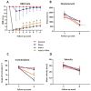
Figure 1
Figure 1
Behavioral tests after spinal cord injury and epidural stimulation. Four groups of animals were used for this purpose, 15 animals of each of the following: control, sham (stimulator implant but without battery), injury group (laminectomy and spinal cord injury through impact), and injury + stim. In (A), BBB score values were recorded weekly over 8 weeks of observation; notably, after the 5th week, a significant difference (p < 0.05) was observed between the injury + stim and injury groups, in favor of the stimulated group. In terms of general displacement in the open field test (B), both injured groups differed from controls but not from each other. In the inclined plane (C), there was a significant difference in performance in favor of the injury + stim group in relation to the injury group (p < 0.05). For velocity (D), no significant difference was observed among groups. * for p < 0.05, *** for p < 0.001.
Figure 2
Figure 2
Microscope image of Hematoxylin and Eosin staining showing the (A) control, (B) sham, (C) injury, and (D) injury + stim groups. Note the (C) globular increase in cell bodies and pronounced cytoplasmatic vacuolization. In (D), changes in cellular cytoarchitecture were also observed, with regions showing vacuolization and globular enlargement of neuronal cell bodies, but these were less pronounced.
Figure 3
Figure 3
Cytoarchitectural changes induced by epidural stimulation. (AD), confocal microscope pictures showing anti-synaptophysin revealed with AlexaFluor 488 (green) and anti-βIII-tubulin revealed with AlexaFLuor 594 (red), in a representative spinal cord axial slice in the control group (A), sham (B), injury group (C), and injury + stim (D) groups. All stainings were processed in parallel and all confocal pictures were taken with the same equipment settings. Note the intense cytoarchitectonic disorganization observed in (C), which is remarkably reversed under epidural stimulation (D) of all slices corresponding to the Z-height lesion epicenter −0.5 mm; all pictures were taken with the same confocal settings to allow for direct comparisons. In (E), estimative numbers of NeuN-positive neuronal nuclei, in terms of total cells per animal; note significantly larger numbers of NeuN+ cells in the injury + stim group compared with the injury group. In (F), βIII-tubulin fluorescence signal intensity was determined with an epifluorescence microscope (background fluorescence intensity subtracted from the total fluorescence in the frame, recorded in arbitrary units a.u.); no difference was seen between the injury + stim and injury groups. In (G), the same quantification was applied for synaptophysin signal intensity, which was significantly greater in the injury + stim group relative to the injury group (p < 0.05). 0 for no difference, * for p < 0.05, *** for p < 0.001.
Figure 4
Figure 4
Representative photomicrographs of Caspase 3 immunostaining on transverse sections of the ventral horn in the spinal cord from control, injury, and injury + stim groups (revealed with Alexa 594). In (A), a representative image from the control group shows a relatively low expression level of Caspase 3. In (B), a representative image from the injury group with pronounced expression throughout the ventral horn is shown; note the morphological and structural changes in the tissue resulting from severe spinal cord injury. In (C), representative images from the injury + stim group exhibit notable immunofluorescence reactions, albeit with lower reactive luminescence compared with the injury group. Panel (D) presents the bar graphs of intensity measurements in terms of means and SEM; note the significantly different Caspase levels in the injury group, which are significantly higher when compared to the stim and control groups. Moreover, no difference in Caspase levels was seen between controls and stim. (* p < 0.05).
Figure 5
Figure 5
Schematic representation of the experiments. In (A), rats were divided into four groups: control group (without spinal cord injury-SCI, n = 15), sham group (implanted with spinal cord stimulation without the battery-SCS, n = 15), injury group (traumatic SCI, n = 15), and injury + stim group (submitted to traumatic SCI and full SCS, n = 15). Seven days post-SCI, the sham and stimulation groups had their spinal cords re-exposed. A paddle electrode and an internal pulse generator were implanted. The device delivered a 30 Hz, 500 μs, 110 mV pulse for 40 min daily over 60 days. In (B), locomotor function was evaluated weekly using the BBB scale, muscle strength was tested using the inclined plane test, and locomotor activity was assessed using the open field test. After the behavioral tasks, the rats were perfused and their spinal cords were processed for histological analysis. In panel (C), the timeline illustrates the sequence of experimental events, including pre-surgical evaluations, weekly analyses, final assessments, animal sacrifice, and immunohistochemical processing.
Figure 6
Figure 6
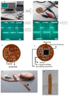
(A,B) bench test. Note the power supply adjusted to 5 V supplying the stimulation device; (C,D): oscilloscopic recording of the output current from the device (110 mV and 30.7 Hz). Figures (E,F) display the first version of the stimulator with its basic components, while Figure (G) depicts the final version of the device, # (Main microchip stimulation); * (Power supply); arrow (implantable electrode); (H) epidural implant electrode. The implantable pulse generator was designed and manufactured in our laboratory. It was conceived as a single piece, covered with dimethylpolysiloxane to avoid a battery leak. The lithium battery Sony CR2032, 220 mAh and 3 V, had enough charge to deliver a quadratic pulse of 30 Hz, 500 μs for 40 min a day over 60 days; the whole system required 1 mA/h. By the time of each animal’s sacrifice, the device was explanted and battery tension stability was assessed by millimeter (always higher than 2.9 V).

Similar articles

Cited by

References

    1. GBD Collaborator Network Global, regional, and national burden of disorders affecting the nervous system, 1990-2021: A systematic analysis for the Global Burden of Disease Study 2021. Lancet Neurol. 2024;23:344–381. doi: 10.1016/S1474-4422(24)00038-3. - DOI - PMC - PubMed
    1. GBD Collaborator Network Global, regional, and national burden of spinal cord injury, 1990–2019: A systematic analysis for the Global Burden of Disease Study 2019. Lancet Neurol. 2023;22:1026–1047. doi: 10.1016/S1474-4422(23)00287-9. - DOI - PMC - PubMed
    1. Hachem L.D., Ahuja C.S., Fehlings M.G. Assessment and management of acute spinal cord injury: From point of injury to rehabilitation. J. Spinal Cord. Med. 2017;40:665–675. doi: 10.1080/10790268.2017.1329076. - DOI - PMC - PubMed
    1. Mariano E.D., Batista C.M., Barbosa B.J., Marie S.K., Teixeira M.J., Morgalla M., Tatagiba M., Li J., Lepski G. Current perspectives in stem cell therapy for spinal cord repair in humans: A review of work from the past 10 years. Arq. Neuropsiquiatr. 2014;72:451–456. doi: 10.1590/0004-282X20140051. - DOI - PubMed
    1. Li J., Lepski G. Cell transplantation for spinal cord injury: A systematic review. Biomed. Res. Int. 2013;2013:786475. doi: 10.1155/2013/786475. - DOI - PMC - PubMed

MeSH terms

LinkOut - more resources