Review
. 2024 Aug 21;25(16):9050.
doi: 10.3390/ijms25169050.

Post-Acute Sequelae and Mitochondrial Aberration in SARS-CoV-2 Infection

Affiliations
Review

Post-Acute Sequelae and Mitochondrial Aberration in SARS-CoV-2 Infection

Charles Ward et al. Int J Mol Sci. .

Abstract

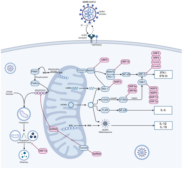
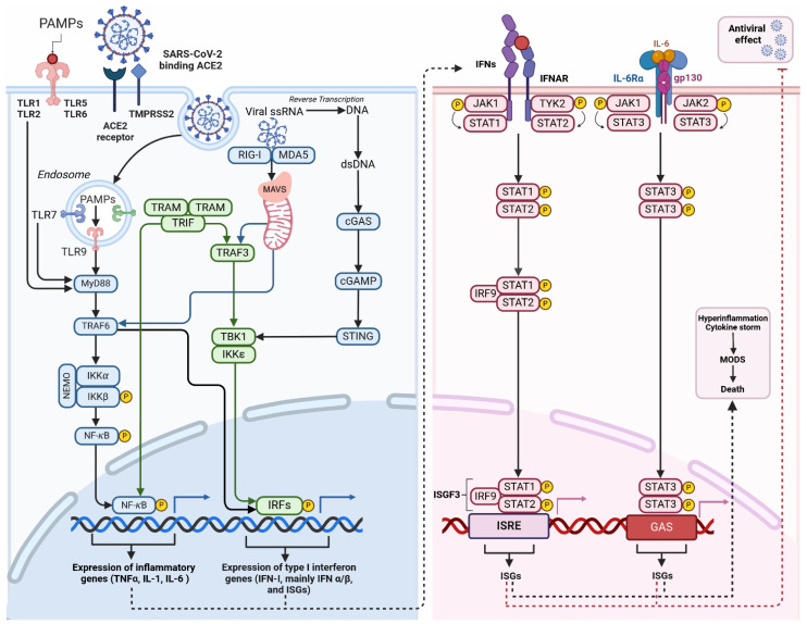
This review investigates links between post-acute sequelae of SARS-CoV-2 infection (PASC), post-infection viral persistence, mitochondrial involvement and aberrant innate immune response and cellular metabolism during SARS-CoV-2 infection. Advancement of proteomic and metabolomic studies now allows deeper investigation of alterations to cellular metabolism, autophagic processes and mitochondrial dysfunction caused by SARS-CoV-2 infection, while computational biology and machine learning have advanced methodologies of predicting virus-host gene and protein interactions. Particular focus is given to the interaction between viral genes and proteins with mitochondrial function and that of the innate immune system. Finally, the authors hypothesise that viral persistence may be a function of mitochondrial involvement in the sequestration of viral genetic material. While further work is necessary to understand the mechanisms definitively, a number of studies now point to the resolution of questions regarding the pathogenesis of PASC.

Keywords: PASC; SARS-CoV-2; autophagy; cell metabolism; innate immunity; long COVID; mitochondria; mitophagy; mtDNA; reactive oxygen species.


Conflict of interest statement

The authors declare no conflicts of interest.

Figures

Figure 1
Figure 1
Mechanism of SARS-CoV-2 entry, mitochondrial damage sensing and innate immune dysregulation. Viral particles enter cells via the angiotensin-converting enzyme 2 (ACE2) receptor and TMPRSS2 protease. In normally functioning cells various mechanisms exist to warn of mitochondrial damage and viral infection, most notably via retinoic acid inducible gene-1 and mitochondrial antivirus signalling protein (RIG-1-MAVS), cyclic GMP–AMP synthase and stimulator of interferon genes (cGAS-STING) signalling, Toll-like receptor-9 (TLR9) and activation of the NOD-, LRR -and pyrin domain containing protein 3 (NLRP3) inflammasome. Damage to mitochondria resulting in the production of ROS initiates mitophagy via the PTEN-induced kinase 1 (PINK1)-Parkin E3 ubiquitin ligase pathway. Infection of cells by SARS-CoV-2 disrupts these axes at a number of control points and causes widespread aberration to systems of innate immunity. Abbreviations: ACE2—angiotensin-converting enzyme 2; ATG9A—autophagy-related protein 9A; cGAMP—cyclic GMP-AMP; cGAS—cyclic GMP-AMP synthase; dsRNA—double-stranded RNA; ETC—electron transport chain; IL-1β—interleukin 1 beta; IL18—interleukin 18; IL-6—interleukin 6; IFN I—type I interferon; IFN III—type III interferon; MAVS—mitochondrial antiviral-signalling protein; NFκB—nuclear factor kappa-light-chain-enhancer of activated B cells; NSP—non-structural protein, ORF—open reading frame; Parkin—E3 ubiquitin-protein ligase parkin; ROS—reactive oxygen species; STING—stimulator of interferon genes; TBK1—TANK-biding kinase 1; TMPRSS2—transmembrane protease, Serine 2; TLR9—Toll-like receptor 9; Tom20—translocase of outer mitochondrial membrane 20; TRAF3—TNF receptor-associated factor 3; mtDNA—mitochondrial DNA. Pink objects indicate material of viral origin, blue objects indicate self-material. (Created with BioRender.com).
Figure 2
Figure 2
Inflammation triggered by macrophage after SARS-CoV-2 infection. Activation of Toll-like receptors (TLRs) occurs when they recognise pathogen-associated molecular patterns (PAMPs). These patterns are typically found in foreign organisms, such as bacteria and viruses. TLR localisation can occur on either the cell surface (TLR-1, -2, -4, -5, -6, -10) or in intracellular compartments such as endosomes (TLR-3, -7, -8, -9). The ability to recognise viral single-stranded RNA (ssRNA) implies its potential for SARS-CoV-2 clearance of SARS-CoV-2. These receptors detect signals and initiate NF-κB and IRF activation. Activation can occur through the MyD88-dependent and MyD88-independent pathways, ultimately leading to the expression of cytokines and interferon (IFN-I). The binding of viral RNA to RIG-I or MDA5 prompts the creation of MAVS polymers in mitochondria, followed by the subsequent attachment of TRAFs. TRAFs activate the NF-κB, and interferon regulatory factors (IRFs), mainly IRF-3 and IRF-7. This process leads to the expression of antiviral interferon-stimulated genes (ISGs) and pro-inflammatory cytokines. Cytosolic DNA sensors, primarily cGAS detect viral DNA, including dsDNA from DNA viruses or reverse-transcribed DNA from retroviruses. Upon binding to dsDNA, cGAS becomes activated and catalyses the production of a second messenger cyclic GMP-AMP (cGAMP). cGAMP binds and activates STING, which recruits and activates TBK1. Activated TBK1 phosphorylates the transcription factor IRF3 leading to the expression of ISGs. Activation of the JAK-STAT pathway by IFN and cytokines initiates the innate immune response against viral infections. The role of this pathway is critical in various physiological and pathological processes like cancer development, inflammation, tissue damage, and viral infections. The binding of Type I IFN to IFNα receptors (IFNAR) activates JAKs, which in turn phosphorylates and moves STATs to the nucleus. This process leads to the expression of antiviral ISGs. Once the STAT1-STAT2 heterodimer is formed, it binds to IRF9 to create the transcriptionally active ISGF3. The early immune response to viral infections heavily relies on the activation of the Jak2/STAT3 signalling pathway by Il-6, which facilitates virus clearance through neutrophils. The IL-6 protein binds to the IL-6R receptor, which is composed of the IL-6α receptor molecule and the gp130 signal transducer, allowing cell signalling. JAK2 activation induced by IL-6 through gp130 results in the activation of STATs, particularly STAT3. Abbreviations: ISGs—interferon-stimulated genes; cGAMP—cyclic GMP-AMP; cGAS—(cyclic GMP-AMP synthase); GAS—interferon-activated site; IFNs—interferons; IKKα—IκB kinase α; IRF9—interferon regulatory factor 9; ISGF3—INF-stimulated gene factor 3; ISGs—interferon-stimulated genes; ISRE—interferon-stimulated response element; JAK—Janus kinase; MDA5—melanoma differentiation-associated protein 5; MODS—multiple organ dysfunction syndrome; MyD88—myeloid differentiation primary response 88; NEMO—NF-κB essential modulator; PAMPs—pathogen-associated molecular patterns; RIG-I—retinoic acid-inducible gene I; STAT—signal transducer and activator of transcription; STING—stimulator of interferon genes; TBK1—TANK-binding kinase; TRAF—tumour necrosis factor receptor-related factor; TRAF6—tumour necrosis factor receptor-related factor 6; TRAM—trif-related adaptor molecule; TRIF—domain-containing adaptor protein inducing interferon β; Tyk2—tyrosine kinase 2. Different colours in the figure represent various components of the signaling pathways–blueindicates MyD88-dependent and cGAS-STING pathways, green represents IRF, and pink represents the JAK/STAT pathway. c. (Created with BioRender.com).

Similar articles

Cited by

References

    1. Post COVID-19 Condition (Long COVID) [(accessed on 30 January 2024)]. Available online: https://www.who.int/europe/news-room/fact-sheets/item/post-covid-19-cond....
    1. Nalbandian A., Sehgal K., Gupta A., Madhavan M.V., McGroder C., Stevens J.S., Cook J.R., Nordvig A.S., Shalev D., Sehrawat T.S., et al. Post-Acute COVID-19 Syndrome. Nat. Med. 2021;27:601–615. doi: 10.1038/s41591-021-01283-z. - DOI - PMC - PubMed
    1. Bell M.L., Catalfamo C.J., Farland L.V., Ernst K.C., Jacobs E.T., Klimentidis Y.C., Jehn M., Pogreba-Brown K. Post-Acute Sequelae of COVID-19 in a Non-Hospitalized Cohort: Results from the Arizona CoVHORT. PLoS ONE. 2021;16:e0254347. doi: 10.1371/journal.pone.0254347. - DOI - PMC - PubMed
    1. Overview|COVID-19 Rapid Guideline: Managing the Long-Term Effects of COVID-19|Guidance|NICE. [(accessed on 30 January 2024)]. Available online: https://www.nice.org.uk/guidance/NG188.
    1. Davis H.E., McCorkell L., Vogel J.M., Topol E.J. Long COVID: Major Findings, Mechanisms and Recommendations. Nat. Rev. Microbiol. 2023;21:133–146. doi: 10.1038/s41579-022-00846-2. - DOI - PMC - PubMed

LinkOut - more resources