. 2024 Aug 21;25(16):9057.
doi: 10.3390/ijms25169057.

AAV-Mediated Expression of miR-17 Enhances Neurite and Axon Regeneration In Vitro

Affiliations

AAV-Mediated Expression of miR-17 Enhances Neurite and Axon Regeneration In Vitro

Raquel Alves Almeida et al. Int J Mol Sci. .

Abstract

Neurodegenerative disorders, including traumatic injuries to the central nervous system (CNS) and neurodegenerative diseases, are characterized by early axonal damage, which does not regenerate in the adult mammalian CNS, leading to permanent neurological deficits. One of the primary causes of the loss of regenerative ability is thought to be a developmental decline in neurons' intrinsic capability for axon growth. Different molecules are involved in the developmental loss of the ability for axon regeneration, including many transcription factors. However, the function of microRNAs (miRNAs), which are also modulators of gene expression, in axon re-growth is still unclear. Among the various miRNAs recently identified with roles in the CNS, miR-17, which is highly expressed during early development, emerges as a promising target to promote axon regeneration. Here, we used adeno-associated viral (AAV) vectors to overexpress miR-17 (AAV.miR-17) in primary cortical neurons and evaluate its effects on neurite and axon regeneration in vitro. Although AAV.miR-17 had no significant effect on neurite outgrowth and arborization, it significantly enhances neurite regeneration after scratch lesion and axon regeneration after axotomy of neurons cultured in microfluidic chambers. Target prediction and functional annotation analyses suggest that miR-17 regulates gene expression associated with autophagy and cell metabolism. Our findings suggest that miR-17 promotes regenerative response and thus could mitigate neurodegenerative effects.

Keywords: axon; damage; miR-17; regeneration.


Conflict of interest statement

The authors declare that they have no conflicts of interest.

Figures

Figure 1
Figure 1
Transduction of primary cortical neurons with AAV.miR-17 and AAV.CTRL. (A) Vector maps of the control AAV (AAV. CTRL) and the AAV to overexpress the miR-17 (AAV.miR-17), both of them express the mCherry fluorophore under the control of a human synapsin (hSyn) promoter. The miR-17 expression is under the control of the H1 promoter. (B) Scheme of experimental setup for testing the transduction of AAV vectors. PREP: day of preparation of E18 rat cortical neurons; DIV: day in vitro; AAV: transduction with AAV vectors; IMG: imaging. (C) Representative images of cortical neurons 8 days after transduction with AAV.CTRL or AAV.miR-17 vectors showing the expression of the mCherry fluorescent protein (red), phase contrast, and the merge. Scale bar: 50 µm. (D) Quantification of cell viability through the MTT assay at the DIV8. (n = 3 independent cultures). Data are presented as single data points and means ± SEM. p = 0.9778, according to a two-tailed unpaired t-test.
Figure 2
Figure 2
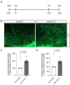
AAV.miR-17 has no effect on neurite outgrowth. (A) Scheme of experimental setup for neurite outgrowth assay. PREP: day of preparation of E18 rat cortical neurons; DIV: day in vitro; AAV: transduction with AAV vectors; IMG: imaging. (B) Representative images of DIV8 cortical neurons transduced with AAV.miR-17 or AAV.CTRL vectors showing the staining for β-III-tubulin (Tuj1, green). Scale bar: 50 μm. (C) The same representative images are shown in (B) after image processing for the quantification of neurite outgrowth. (D) Quantification of the area of the Tuj1 staining normalized by the number of cell bodies. (n = 6 independent cultures). Data are presented as single data points and means ± SEM. p = 0.7907, according to a two-tailed unpaired t-test.
Figure 3
Figure 3
The complexity of the neurite arborization is not influenced by AAV.miR-17. (A) Scheme of the experimental setup for neurite arborization complexity assay. PREP: day of preparation of E18 rat cortical neurons; DIV: day in vitro; AAV: transduction with AAV vectors; IMG: imaging. (B) Representative images of DIV8 isolated cortical neurons transduced with AAV.miR-17 or AAV.CTRL vectors showing the staining for β-III-tubulin (Tuj1, green). Scale bar: 20 μm. (C) The same representative images are shown in (B) after image processing for the quantification of Sholl analysis, showing the concentric circles and the intersection between them and the neurites. (D) Quantification of the number of intersections between the neurites and the concentric circles at the given radius. Data are presented as means ± SEM (n = 5 independent cultures). (E) Quantification of the total number of intersections between the neurites and all the concentric circles. (n = 5 independent cultures). Data are presented as single data points and means ± SEM. p = 0.1697, according to a two-tailed unpaired t-test.
Figure 4
Figure 4
AAV.miR-17 enhances neurite regeneration after scratch lesions. (A) Scheme of the experimental setup for neurite regeneration assay. PREP: day of preparation of E18 rat cortical neurons; DIV: day in vitro; AAV: transduction with AAV vectors; LES: scratch lesion; IMG: imaging. (B) Representative images of cortical neurons transduced with AAV.CTRL or AAV.miR-17 vector and 24 h after the scratch lesion showing the regenerating neurites stained for β-III-tubulin (Tuj1, green). The dashed line indicates the scratch lesion border. Scale bar: 50 μm. (C) Quantification of the neurite length in the lesion area in a distance from 100 µm to 200 µm from the scratch border, normalized by the control group (AAV.CTRL). (D) Quantification of the number of neurites that re-growth until the distance of 200 µm from the scratch border, normalized by the control group (AAV.CTRL). (n = 5 independent cultures). Data are presented as single data points and means ± SEM. Indicated p-value according to one-sample t-test.
Figure 5
Figure 5
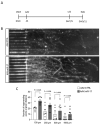
AAV.miR-17 increases axonal regeneration after axotomy. (A) Scheme of experimental setup for axonal regeneration analysis in microfluidic chambers. PREP: day of preparation of E18 rat cortical neurons; DIV: day in vitro; AAV: transduction with AAV vectors; LES: axotomy; IMG: imaging. (B) Representative images of cortical neurons transduced with AAV.CTRL or AAV.miR-17 vector and seeded in microfluidic chambers 48 h after the axotomy, showing the regenerating axons labeled with the mCherry fluorescent protein (white). Scale bar: 200 μm. (C) Quantification of the number of axons that re-growth until the given distances from the microgroove exit. (n = 7 independent cultures). Data are presented as single data points and means ± SEM. Indicated p-value according to two-tailed unpaired t-test.
Figure 6
Figure 6
Enrichment analyses for the targets of hsa-miR-17-5p. (A) Target prediction results reveal a total of 579 predicted targets for hsa-miR-17-5p—of which 252 have been experimentally validated. The list of validated targets used for all enrichment analyses is presented here. (BD) Gene ontology analyses for the targets of hsa-miR-17-5p. These analyses were divided into sub-categories: Biological Processes (B), Cellular Component (C), and Molecular Function (D). (E) Protein-protein interaction (PPI) networks for the targets of hsa-miR-17-5p that composed GO-BP enrichment results. STAT3, MAPK1, CREB1, E2F3/5, MCL1, CCND1/2, and CDKN1A appear as important molecular hubs.

Similar articles

References

    1. Neukomm L.J., Freeman M.R. Diverse cellular and molecular modes of axon degeneration. Cell Press. 2014;24:515–523. doi: 10.1016/j.tcb.2014.04.003. - DOI - PMC - PubMed
    1. Goldberg J.L., Klassen M.P., Hua Y., Barres B.A. Amacrine-Signaled Loss of Intrinsic Axon Growth Ability by Retinal Ganglion Cells. Science. 2002;296:1860–1864. doi: 10.1126/science.1068428. - DOI - PubMed
    1. Yiu G., He Z. Glial inhibition of CNS axon regeneration. Nature. 2006;7:617–627. doi: 10.1038/nrn1956. - DOI - PMC - PubMed
    1. Bouslama-Oueghlani L., Wehrle R., Sotelo C., Dusart I. The Developmental Loss of the Ability of Purkinje Cells to Regenerate Their Axons Occurs in the Absence of Myelin: Na In Vitro Model to Prevent Myelination. J. Neurosci. 2003;23:8318–8329. doi: 10.1523/JNEUROSCI.23-23-08318.2003. - DOI - PMC - PubMed
    1. Blackmore M., Letourneau P.C. Changes within Maturing Neurons Limit Axonal Regeneration in the Developing Spinal Cord. J. Neurobiol. 2006;66:348–360. doi: 10.1002/neu.20224. - DOI - PubMed

LinkOut - more resources