. 2024 Aug 21;25(16):9061.
doi: 10.3390/ijms25169061.

tRF-Gly-GCC in Atretic Follicles Promotes Ferroptosis in Granulosa Cells by Down-Regulating MAPK1

Affiliations

tRF-Gly-GCC in Atretic Follicles Promotes Ferroptosis in Granulosa Cells by Down-Regulating MAPK1

Yuheng Pan et al. Int J Mol Sci. .

Abstract

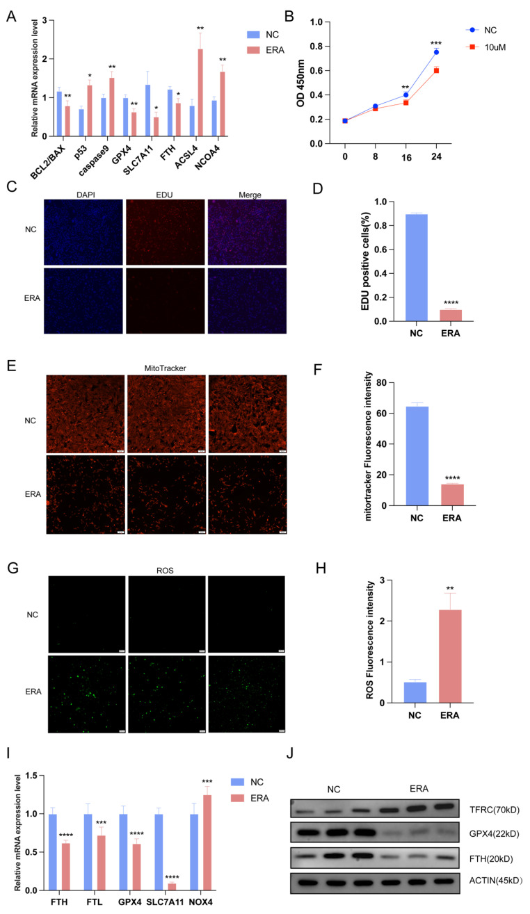
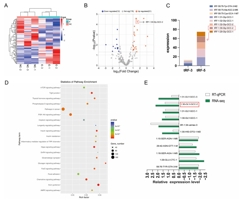
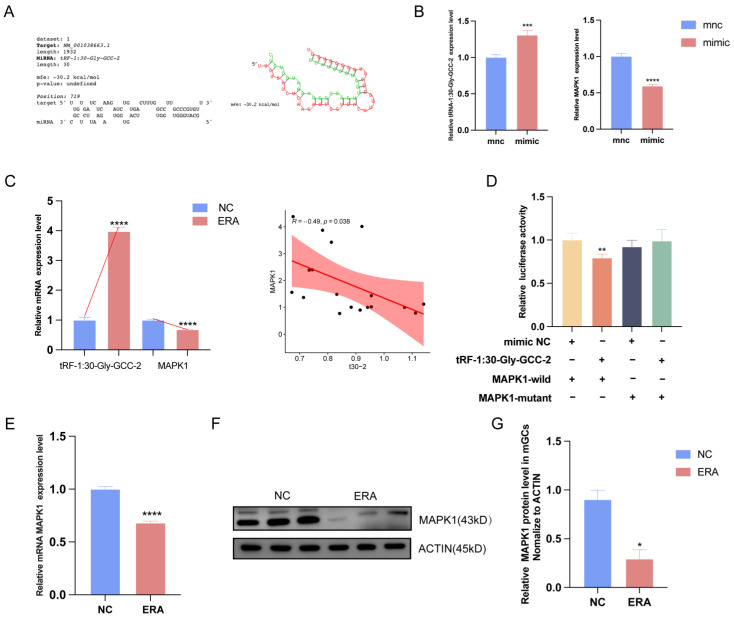
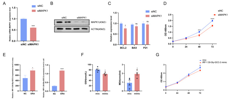
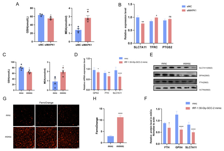
Follicle development refers to the process in which the follicles in the ovary gradually develop from the primary stage to a mature state, and most primary follicles fail to develop normally, without forming a dense granular cell layer and cell wall, which is identified as atretic follicles. Granulosa cells assist follicle development by producing hormones and providing support, and interference in the interaction between granulosa cells and oocytes may lead to the formation of atretic follicles. Ferroptosis, as a non-apoptotic form of death, is caused by cells accumulating lethal levels of iron-dependent phospholipid peroxides. Healthy follicles ranging from 4 to 5 mm were randomly divided into two groups: a control group (DMSO) and treatment group (10 uM of ferroptosis inducer erastin). Each group was sequenced after three repeated cultures for 24 h. We found that ferroptosis was associated with atretic follicles and that the in vitro treatment of healthy follicles with the ferroptosis inducer erastin produced a phenotype similar to that of atretic follicles. Overall, our study elucidates that tRF-1:30-Gly-GCC-2 is involved in the apoptosis and ferroptosis of GCs. Mechanistically, tRF-1:30-Gly-GCC-2 inhibits granulosa cell proliferation and promotes ferroptosis by inhibiting Mitogen-activated protein kinase 1 (MAPK1). tRF-1:30-Gly-GCC-2 may be a novel molecular target for improving the development of atretic follicles in ovarian dysfunction. In conclusion, our study provides a new perspective on the pathogenesis of granulosa cell dysfunction and follicular atresia.

Keywords: MAPK1; atretic follicle; ferroptosis; granular cell; tRF-1:30-Gly-GCC-2.


Conflict of interest statement

The authors declare no conflicts of interest.

Figures

Figure 1
Figure 1
Ferroptosis occurs in atretic follicles. (A) Comparison of phenotypes between healthy follicles and atretic follicles. (B) The content of GSH, MDA, and Fe2+ in healthy and atretic follicles. (C) To further confirm the effect of ferroptosis on follicular development, we detected the down-regulated standard genes FTH1, FTL, and SLC7A11 and the up-regulated marker genes PTGS2 and ACSL4 in follicles by qRT-PCR. Data represented the mean ± SEM of at least three biological replicates. * p < 0.05, ** p < 0.01, *** p < 0.001 and **** p < 0.0001, compared to control.
Figure 2
Figure 2
Ferroptosis inducers induced follicular atresia and granular cell ferroptosis in erastin. (A) The expression of ferroptosis marker genes were detected by qRT-PCR in erastin-treated follicles. The result of CCK-8 (B) is consistent with the result of EDU experiment (C,D). Mitotracker (50 μm) staining is used to detect mitochondrial activity in control group and erastin group (E,F); ROS (200 μm) staining is used to detect reactive oxygen species in control group and erastin group (G,H). Erastin induced ferroptosis in granular cells, and qRT-PCR (I) results are consistent with WB (J). Data represented the mean ± SEM of at least three biological replicates. * p < 0.05, ** p < 0.01, *** p < 0.001 and **** p < 0.0001, compared to control.
Figure 3
Figure 3
tRFs number statistics and clustering heat maps of differences in sequencing between healthy follicles and inducer-treated follicles. (A) tRFs clustering heat map of sequencing data differences. (B) Sequencing data variance tRFs volcano map. (C) The abundance values of tRF-3 and tRF-5 were analyzed. (D) KEGG enrichment analysis. (E) qRT-PCR results verified the sequencing data.
Figure 4
Figure 4
MAPK1 is the target of tRF-1:30-Gly-GCC-2. (A) The binding site of tRF-1:30-Gly-GCC-2 to MAPK1 was predicted. (B) The expression of MAPK1 in control group and overexpression group. (C) Correlation analysis of tRF and MAPK1. (D) Dual luciferase reports detected tRF-1:30-Gly-GCC-2 binding to MAPK1. (E) The expression level of MAPK1 in the control group and erastin group was detected by qRT-PCR, and the results of WB (F,G) were consistent with them. Data represented the mean ± SEM of at least three biological replicates. * p < 0.05, ** p < 0.01, *** p < 0.001 and **** p < 0.0001, compared to control.
Figure 5
Figure 5
Overexpression of tRF-1:30-Gly-GCC-2 or MAPK1 knockdown inhibited granulosa cell proliferation. (A) The expression of MAPK1 in siNC group and siMAPK1 group. The results of WB (B) are consistent with those of PCR. The expression of tRF-1:30-Gly-GCC-2 was observed in follicular and granulosa cells and erastin group. (C) Apoptosis marker genes in siMAPK1 group in granulosa cells were detected by qRT-PCR. (D) CCK-8 detected the viability of cell knockdown MAPK1. (E) Expression of tRF-1:30-Gly-GCC-2 in follicle and granulosa cells treated with erastin. (F) Contents of GSH and MDA in mnc group and tRF-1:30-Gly-GCC-2mimic group. (G) CCK-8 detected the viability of cells overexpressing tRF-1:30-Gly-GCC-2. Data represented the mean ± SEM of at least three biological replicates. ns > 0.05, * p < 0.05, ** p < 0.01, *** p < 0.001 and **** p < 0.0001, compared to control.
Figure 6
Figure 6
tRF-1:30-Gly-GCC-2 promotes ferroptosis in granular cells through MAPK1. (A) The contents of GSH and MDA in cells after transfection with siMAPK1. (B) The expression of iron sag marker genes SLC7A11, TFRC, and PTGS2 after MAPK1 knockout was detected by qRT-PCR. (C) Contents of GSH and MDA in mnc group and tRF-1:30-Gly-GCC-2mimic group. (D) The expression of ferroptosis marker gene GPX4, FTH, and SLC7A11 after overexpression of tRF-1:30-Gly-GCC-2 was detected by qRT-PCR. The results of WB (E,F) were consistent with them. (G,H) FerroOrange (200 μm) content of iron ion probe in control group and overexpression group. Data represented the mean ± SEM of at least three biological replicates. ns > 0.05, * p < 0.05, ** p < 0.01 and **** p < 0.0001, compared to control.

Similar articles

Cited by

References

    1. Beltran-Alcrudo D., Falco J.R., Raizman E. Transboundary spread of pig diseases: The role of international trade and travel. BMC Vet. Res. 2019;15:64. doi: 10.1186/s12917-019-1800-5. - DOI - PMC - PubMed
    1. Stringer J.M., Alesi L.R., Winship A.L. Beyond apoptosis: Evidence of other regulated cell death pathways in the ovary throughout development and life. Hum. Reprod. Update. 2023;29:434–456. doi: 10.1093/humupd/dmad005. - DOI - PMC - PubMed
    1. Chang C.-W., Sung Y.-W., Hsueh Y.-W. Growth hormone in fertility and infertility: Mechanisms of action and clinical applications. Front. Endocrinol. 2022;13:1040503. doi: 10.3389/fendo.2022.1040503. - DOI - PMC - PubMed
    1. Fan X., Bialecka M., Moustakas I. Single-cell reconstruction of follicular remodeling in the human adult ovary. Nat. Commun. 2019;10:3164. doi: 10.1038/s41467-019-11036-9. - DOI - PMC - PubMed
    1. Gougeon A. Some aspects of the dynamics of ovarian follicular growth in the human. Acta Eur. Fertil. 1989;20:185–192. - PubMed

Substances

LinkOut - more resources