. 2024 Aug 21;25(16):9063.
doi: 10.3390/ijms25169063.

Characterization and Functional Analysis of the 17-Beta Hydroxysteroid Dehydrogenase 2 (hsd17b2) Gene during Sex Reversal in the Ricefield Eel (Monopterus albus)

Affiliations

Characterization and Functional Analysis of the 17-Beta Hydroxysteroid Dehydrogenase 2 (hsd17b2) Gene during Sex Reversal in the Ricefield Eel (Monopterus albus)

Ruyi Chen et al. Int J Mol Sci. .

Abstract

In mammals, 17-beta hydroxysteroid dehydrogenase 2 (Hsd17b2) enzyme specifically catalyzes the oxidation of the C17 hydroxyl group and efficiently regulates the activities of estrogens and androgens to prevent diseases induced by hormone disorders. However, the functions of the hsd17b2 gene involved in animal sex differentiation are still largely unclear. The ricefield eel (Monopterus albus), a protogynous hermaphroditic fish with a small genome size (2n = 24), is usually used as an ideal model to study the mechanism of sex differentiation in vertebrates. Therefore, in this study, hsd17b2 gene cDNA was cloned and its mRNA expression profiles were determined in the ricefield eel. The cloned cDNA fragment of hsd17b2 was 1230 bp, including an open reading frame of 1107 bp, encoding 368 amino acid residues with conserved catalytic subunits. Moreover, real-time quantitative reverse transcription polymerase chain reaction (RT-qPCR) analysis showed that hsd17b2 mRNA expressed strongly in the ovaries at early developmental stages, weakly in liver and intestine, and barely in testis and other tissues. In particular, hsd17b2 mRNA expression was found to peak in ovaries of young fish and ovotestis at the early stage, and eventually declined in gonads from the late ovotestis to testis. Likewise, chemical in situ hybridization results indicated that the hsd17b2 mRNA signals were primarily detected in the cytoplasm of oogonia and oocytes at stage I-II, subsequently concentrated in the granulosa cells around the oocytes at stage Ⅲ-Ⅳ, but undetectable in mature oocytes and male germ cells. Intriguingly, in ricefield eel ovaries, hsd17b2 mRNA expression could be significantly reduced by 17β-estradiol (E2) or tamoxifen (17β-estradiol inhibitor, E2I) induction at a low concentration (10 ng/mL) and increased by E2I induction at a high concentration (100 ng/mL). On the other hand, both the melatonin (MT) and flutamide (androgen inhibitor, AI) induction could significantly decrease hsd17b2 mRNA expression in the ovary of ricefield eel. This study provides a clue for demonstrating the mechanism of sexual differentiation in fish. The findings of our study imply that the hsd17b2 gene could be a key regulator in sexual differentiation and modulate sex reversal in the ricefield eel and other hermaphroditic fishes.

Keywords: chemical in situ hybridization; hormone induction; hsd17b2; ricefield eel; sex reversal.


Conflict of interest statement

The authors declare no conflicts of interest.

Figures
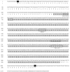
Figure 1
Figure 1
Identification of hsd17b2 cDNA in Monopterus albus. The initiation codon of ATG and stop codon TGA are highlighted in black. The conserved structure of the SDR superfamily, i.e., the Rossmann-folding domain, is highlighted in gray. The classical binding motifs of 17 beta-HSD are in frames, namely TGxxxGxG, YxxxK, PGxxxT, and NNAG. * below the ‘TGA’ for the stop codon.
Figure 2
Figure 2
Homologous analysis and phylogenetic tree of Hsd17b2 proteins in Monopterus albus. (A) Multiple alignment of amino acid (aa) sequences of Hsd17b2 proteins. The protein sequences retrieved from NCBI (https://www.ncbi.nlm.nih.gov/, accessed on 16 May 2023), were aligned and calculated based on a ClustalW algorithm in DNAMAN (https://www.lynnon.com/dnaman.html, accessed on 16 May 2023). The amino acid sequence with 100% identity is highlighted in black, sequences with 75–100% identity are in gray, and sequences with 50% identity are in light gray. The TMD transmembrane region, the Rossmann-folding domain, and low complexity region are highlighted with black frames respectively. The species names and homology are listed at the end of the sequences. (B) Phylogenetic tree of Hsd17b2 proteins. The neighbor-joining phylogenetic tree of Hsd17b2 proteins was constructed by MEGA version 7 (https://www.megasoftware.net/, accessed on 16 May 2023), with a set of 1000 bootstraps in the neighbor-joining method. Scale bar tagged with 0.10 indicates the genetic distance, the number on each branch represents the bootstrap value.
Figure 3
Figure 3
Bioinformatics analysis of Hsd17b2 protein in Monopterus albus. (A) Ribbon diagram of Hsd17b2 protein, containing α-helices and β-strands. α-helices are shown as coiled ribbons and β-strands as arrows; lines indicate random coils. (B) Predicted transmembrane region; the ordinate represents the probability of transmembrane transfer, and the abscissa indicates the sites of amino acid residues. (C) Hydrophilicity map of Hsd17b2 protein; the ordinate indicates the hydrophilicity index ranging from −2.711 to 2.367. (D) Signal peptide predicted by SignalP 5.0 (https://services.healthtech.dtu.dk/services/SignalP-5.0/, accessed on 16 May 2023). The probability of signal protein was about 5.98% and located at residues 1-21.
Figure 4
Figure 4
RT-qPCR analysis of zar1 and hsd17b2 mRNA in tissues. (A,B) Tissue-specific analysis of zar1 and hsd17b2 mRNA in adult M. albus tissues. A panel of tissues was collected and examined in this study, including the intestine (In), brain (Br), heart (He), liver (Li), spleen (Sp), kidney (Ki), testis (Te), and ovary (Ov). (C) Expression profiles of hsd17b2 mRNA in gonads at different developmental stages. (D) Hematoxylin and eosin (HE) staining shows the structures of gonads at different developmental stages examined in this study. Ov1, ovaries from juvenile fish; Ov2, ovaries from young fish; Ov3, ovaries from adult fish; OT1, ovotestis at the early stage; OT2, ovotestis at the middle stage; Te, testis; scale bars, 200 μm. In (AC), different letters (a–d) represent the significance between groups.
Figure 5
Figure 5
Cellular distribution of zar1 and hsd17b2 mRNA in ovaries. Chemical in situ hybridization was conducted on paraffin-embedded sections of ovaries. Antisense and sense RNA probes of hsd17b2 gene were labeled with DIG; signals were developed with NBT/BCIP (in purple). (AC) Ovaries from intersexual individuals; (DF) ovaries from juvenile individuals; (GI) ovaries from young fish. Cellular distribution results indicated that hsd17b2 mRNA was strongly expressed in oogonia and then shifted into the somatic cells wrapping up the oocytes filled with yolk. The zar1 gene specifically expressed in oocytes was used as the control for analyzing the cellular distribution of hsd17b2 mRNA in the gonads of ovotestis at the early stage. I–IV, IIIa, IIIb, represents oocytes at different developmental stages; BV, blood vessels; DO, degenerated oocytes; GL, gonadal lamellae.
Figure 6
Figure 6
Cellular distribution of hsd17b2 mRNA in ovotestis and testis. (AC) Ovotestis at the early stage; (DF) ovotestis at the late stage; (GI) mature testis. The CISH showed that hsd17b2 mRNA expression patterns in gonads at the ovotestis stages were similar to those in ovaries. However, no signal was detected in the gonads of ovotestis at the late stage, and in testis, the testicular cell nuclei were counterstained by propidium iodide (PI, in red). I–IV, Ⅲa, Ⅲb represent oocytes at the different developmental stages. Sc1, primary spermatocyte; Sc2, secondary spermatocyte; Spd, spermatids; Sp, sperm; BV, blood vessels; DO, degenerated oocytes; GL, gonadal lamellae; MC, mesenchyme cluster.
Figure 7
Figure 7
Effects of sex hormone treatment on hsd17b2 mRNA expression in the Monopterus albus ovary. The ovaries of young fish were dissected and treated with 17β-estradiol (E2), 17β-estradiol inhibitor (E2I), melatonin (MT), and flutamide (androgen inhibitor, AI) respectively, and hsd17b2 mRNA expression in the treated ovaries was examined by RT-qPCR. (A) Treatment with E2 and E2I at 10 ng/mL and 100 ng/mL. (B) MT and AI at 10 ng/mL and 100 ng/mL. The group treated with 0.1% DMSO was set as the control. Each treatment group was set up in triplicates, and the experiment was repeated twice. Data are shown as means ± SEM (n = 3). **, extremely significant (p < 0.01); ns, no significance (p > 0.05).

Similar articles

References

    1. Kallberg Y., Oppermann U., Jörnvall H., Persson B. Short-chain dehydrogenases/reductases (SDRs) Eur. J. Biochem. 2002;269:4409–4417. doi: 10.1046/j.1432-1033.2002.03130.x. - DOI - PubMed
    1. Peltoketo H., Luu-The V., Simard J., Adamski J. 17beta-hydroxysteroid dehydrogenase (HSD)/17-ketosteroid reductase (KSR) family; nomenclature and main characteristics of the 17HSD/KSR enzymes. J. Mol. Endocrinol. 1999;23:1–11. doi: 10.1677/jme.0.0230001. - DOI - PubMed
    1. Boucher E., Provost P., Plante J., Tremblay Y. Androgen receptor and 17beta-HSD type 2 regulation in neonatal mouse lung development. Mol. Cell Endocrinol. 2009;311:109–119. doi: 10.1016/j.mce.2009.06.012. - DOI - PubMed
    1. Labrie F., Luu-The V., Lin S., Simard J., Labrie C., El-Alfy M., Pelletier G., Bélanger A. Intracrinology: Role of the family of 17 beta-hydroxysteroid dehydrogenases in human physiology and disease. J. Mol. Endocrinol. 2000;25:1–16. doi: 10.1677/jme.0.0250001. - DOI - PubMed
    1. Lukacik P., Kavanagh K., Oppermann U. Structure and function of human 17beta-hydroxysteroid dehydrogenases. Mol. Cell. Endocrinol. 2006;248:61–71. doi: 10.1016/j.mce.2005.12.007. - DOI - PubMed

MeSH terms

Substances

LinkOut - more resources