. 2024 Aug 21;25(16):9073.
doi: 10.3390/ijms25169073.

Oxysterol Induces Expression of 60 kDa Chaperone Protein on Cell Surface of Microglia

Affiliations

Oxysterol Induces Expression of 60 kDa Chaperone Protein on Cell Surface of Microglia

Koanhoi Kim et al. Int J Mol Sci. .

Abstract

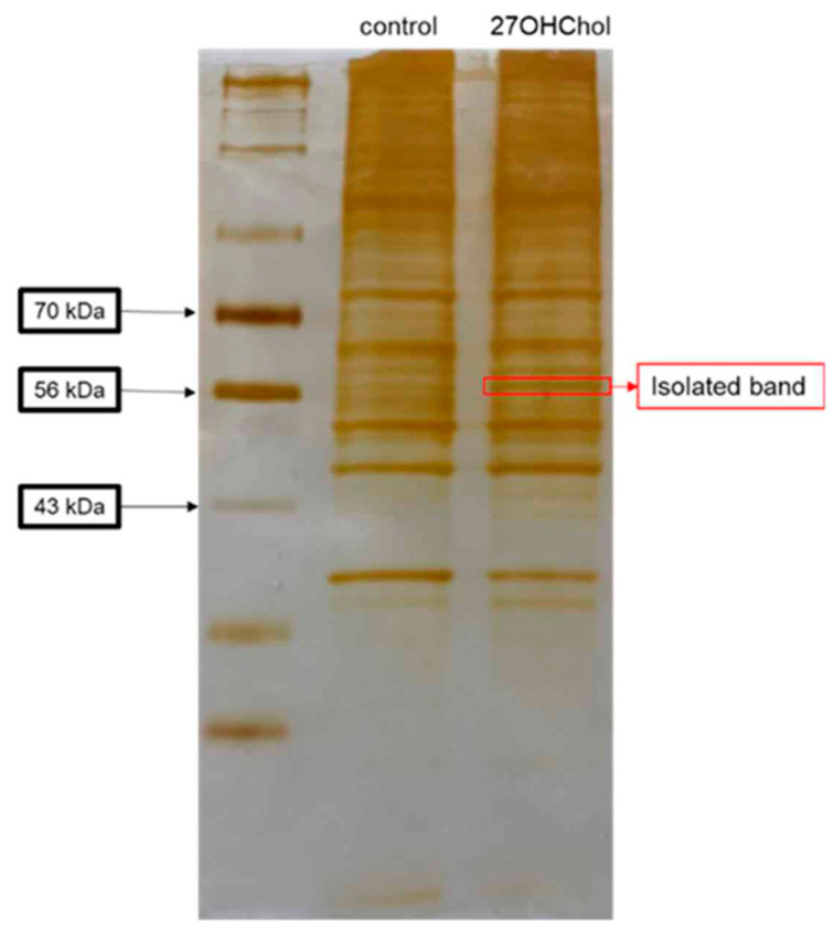
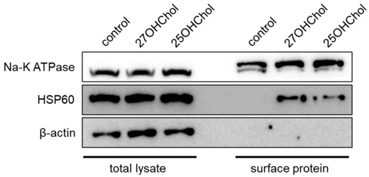
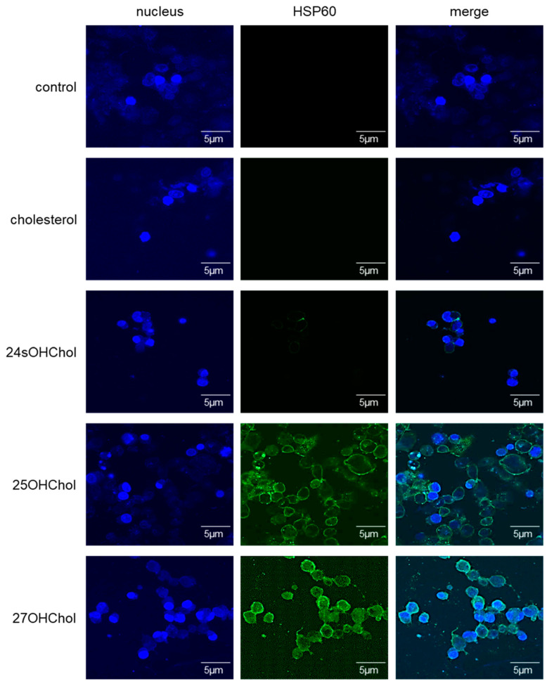
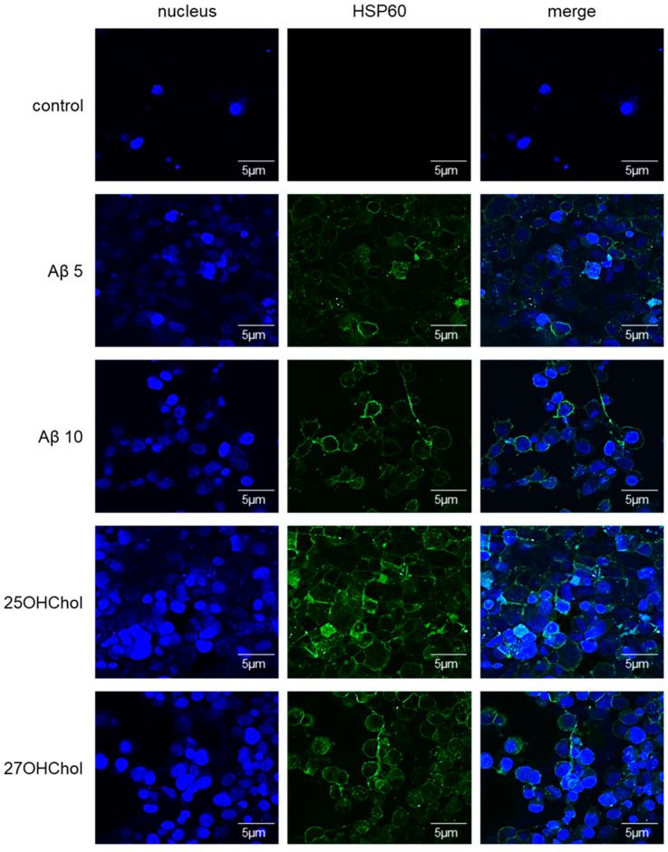
Microglia, essential immune cells in the brain, play crucial roles in neuroinflammation by performing various functions such as neurogenesis, synaptic pruning, and pathogen defense. These cells are activated by inflammatory factors like β-amyloid (Aβ) and oxysterols, leading to morphological and functional changes, including the secretion of inflammatory cytokines and the upregulation of MHC class II molecules. This study focused on identifying specific markers for microglial activation, with a particular emphasis on the roles of oxysterols in this process. We used the HMC3 human microglial cell line to investigate the induction of heat shock protein 60 (HSP60), a chaperonin protein by oxysterols, specifically in the presence of 25-hydroxycholesterol (25OHChol) and 27-hydroxycholesterol (27OHChol). Our findings obtained by the proteomics approach revealed that these oxysterols significantly increased HSP60 expression on microglial cells. This induction was further confirmed using Western blot analysis and immunofluorescence microscopy. Additionally, Aβ1-42 also promoted HSP60 expression, indicating its role as a microglial activator. HSP60 involved in protein folding and immune modulation was identified as a potential marker for microglial activation. This study underscores the importance of HSP60 in the inflammatory response of microglia, suggesting its utility as a target for new therapeutic approaches in neuroinflammatory diseases such as Alzheimer's disease (AD).

Keywords: 25-hydroxycholesterol; 27-hydroxycholesterol; HSP60; microglia; neuroinflammation.


Conflict of interest statement

The authors declare no conflicts of interest.

Figures

Figure 1
Figure 1
Isolated protein band from silver-stained gel. HMC3 cells (1 × 10⁶ cells) were stimulated with or without 1 μg/mL of 27OHChol (concentration of stock: 2 mg/mL) for 48 h, and then surface proteins were isolated with PIERCE® Cell Surface Protein Isolation Kit. The proteins were separated in 10% SDS-contained polyacrylamide gel, and then the gel was stained with PIERCE® Silver Stain Kit. A band was isolated from the gel (red square) and analyzed by MALDI-TOF-MS/MS.
Figure 2
Figure 2
Expression of HSP60 on microglia treated with the oxysterols. HMC3 cells (1 × 10⁶ cells) were treated with 1 μg/mL of 25OHChol and 27OHChol (concentration of stocks: 2 mg/mL) for 48 h. Cell surface proteins harvested from the cells were analyzed with Western blotting. Na-K ATPase is a control for cell surface protein, and β-actin is a loading control. The presented data are representative of three independent experiments.
Figure 3
Figure 3
Visualization of the HSP60 expression on microglia treated with the oxysterols. HMC3 cells seeded on 0.2% gelatin-coated coverslip were stimulated with 1 μg/mL of cholesterol, 24sOHChol, 25OHChol, and 27OHChol (concentration of stocks: 2 mg/mL) for 48 h. The cells were incubated with antibody against HSP60, followed by fluorescence-conjugated 2nd antibody. The samples were visualized with confocal microscopy. The presented data are representative of three independent experiments.
Figure 4
Figure 4
Visualization of the HSP60 expression on microglia treated with Aβ1–42. The cells seeded on 0.2% gelatin-coated coverslip were stimulated with 5 or 10 μM of Aβ1–42 and 1 μg/mL of 25-/27OHChol (concentration of stocks: 2 mg/mL) for 48 h. The cells were incubated with antibody against HSP60, followed by fluorescence-conjugated 2nd antibody. The samples were visualized with confocal microscopy. The presented data are representative of three independent experiments.

Similar articles

Cited by

References

    1. Lehnardt S. Innate immunity and neuroinflammation in the CNS: The role of microglia in Toll-like receptor-mediated neuronal injury. Glia. 2009;58:253–263. doi: 10.1002/glia.20928. - DOI - PubMed
    1. Casano A.M., Peri F. Microglia: Multitasking Specialists of the Brain. Dev. Cell. 2015;32:469–477. doi: 10.1016/j.devcel.2015.01.018. - DOI - PubMed
    1. Thameem Dheen S., Kaur C., Ling E.-A. Microglial Activation and its Implications in the Brain Diseases. Curr. Med. Chem. 2007;14:1189–1197. doi: 10.2174/092986707780597961. - DOI - PubMed
    1. Kim S.U., de Vellis J. Microglia in health and disease. J. Neurosci. Res. 2005;81:302–313. doi: 10.1002/jnr.20562. - DOI - PubMed
    1. Streit W.J., Mrak R.E., Griffin W.S.T. Microglia and neuroinflammation: A pathological perspective. J. Neuroinflamm. 2004;1:14. doi: 10.1186/1742-2094-1-14. - DOI - PMC - PubMed

LinkOut - more resources