. 2024 Aug 21;25(16):9083.
doi: 10.3390/ijms25169083.

Aloperine Inhibits ASFV via Regulating PRLR/JAK2 Signaling Pathway In Vitro

Affiliations

Aloperine Inhibits ASFV via Regulating PRLR/JAK2 Signaling Pathway In Vitro

Renhao Geng et al. Int J Mol Sci. .

Abstract

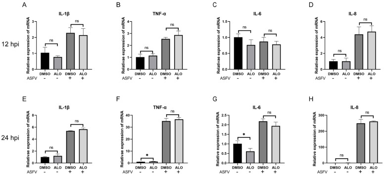
African swine fever (ASF) has become a global pandemic due to inadequate prevention and control measures, posing a significant threat to the swine industry. Despite the approval of a single vaccine in Vietnam, no antiviral drugs against the ASF virus (ASFV) are currently available. Aloperine (ALO), a quinolizidine alkaloid extracted from the seeds and leaves of bitter beans, exhibits various biological functions, including anti-inflammatory, anti-cancer, and antiviral activities. In this study, we found that ALO could inhibit ASFV replication in MA-104, PK-15, 3D4/21, and WSL cells in a dose-dependent manner without cytotoxicity at 100 μM. Furthermore, it was verified that ALO acted on the co- and post-infection stages of ASFV by time-of-addition assay, and inhibited viral internalization rather than directly inactivating the virus. Notably, RT-qPCR analysis indicated that ALO did not exert anti-inflammatory activity during ASFV infection. Additionally, gene ontology (GO) and KEGG pathway enrichment analyses of transcriptomic data revealed that ALO could inhibit ASFV replication via the PRLR/JAK2 signaling pathway. Together, these findings suggest that ALO effectively inhibits ASFV replication in vitro and provides a potential new target for developing anti-ASFV drugs.

Keywords: African swine fever virus; JAK2 signaling pathway; PRLR; aloperine; transcriptomics.


Conflict of interest statement

The authors declare no conflicts of interest.

Figures

Figure 1
Figure 1
ALO inhibited ASFV replication in vitro. (A) Chemical structure of ALO. (B) The cytotoxicity of ALO in MA-104 cells was evaluated by CCK-8 assay after treatment for 48 h. (CE) ASFV-infected MA-104 cells were treated with different concentrations of ALO (2.5, 5, 10, or 20 μM) for 24 h. The effect of ALO anti-ASFV was evaluated by fluorescence intensity, scale bar = 100 μm (C). The protein level of p72 and p30 was examined by Western blot (D), and the viral gene A137R copies (E) were determined by qPCR. (F) Viral gene A137R copies were detected by qPCR in ASFV-infected cells treated with ALO at different time points (24, 48, and 72 hpi). (GI) The cytotoxicity of ALO in PK-15, 3D4/21, and WSL cells was evaluated by CCK-8 assay after treatment for 48 h. (JL) The effect of ALO against ASFV in PK-15, 3D4/21, and WSL cells was determined by RT-qPCR. The data obtained from three independent experiments was analyzed using GraphPad Prism 8.0. Data. *** p < 0.001 and ns p > 0.05, compared to the DMSO control, respectively.
Figure 2
Figure 2
The inhibition stages of ALO on ASFV. (A) The schematic diagram for the process of ALO treatment at different stages of ASFV infection. (B) MA-104 cells were treated with ALO pre-, co-, or post-infection of ASFV. The samples were collected at 24 hpi and evaluated by qPCR and Western blot assay. (C) Effect of ALO treatment on the ASFV entry stage including attachment and internalization was determined by qPCR and Western blot assay. (D) Effect of ALO on ASFV inactivation after being treated for 1 h and 3 h, respectively, were determined by qPCR and Western blot assay. The samples were collected at 24 hpi and evaluated by qPCR and Western blot assay. * p < 0.05, *** p < 0.001, and ns p > 0.05, compared to the DMSO control, respectively.
Figure 3
Figure 3
Effect of ALO on inflammatory cytokines. (AD) RT-qPCR analysis of ASFV-infected MA-104 cells treated with 20 μM ALO; samples were collected at 12 hpi. (EH) RT-qPCR analysis of ASFV-infected MA-104 cells treated with 20 μM ALO; samples were collected at 24 hpi. * p < 0.05 and ns p > 0.05 compared to the DMSO control, respectively.
Figure 4
Figure 4
DEGs in MA-104 cells infected with ASFV treated with/without ALO. (A) The number of significantly up-regulated DEGs (log2FC ≥ 1 and q < 0.05) and down-regulated DEGs (log2FC ≤ −1 and q < 0.05) in each comparison group were counted. The most significant DEGs (top 20) in the groups of ASFV vs. Mock (B), ASFV + ALO vs. Mock (C), and ASFV + ALO vs. ASFV (D) were shown in the volcano maps.
Figure 5
Figure 5
Gene Ontology (GO) terms enrichment of DEGs. The most significant enriched GO terms (top 50) among the DEGs in the groups of ASFV vs. Mock (A), ASFV + ALO vs. Mock (B), and ASFV + ALO vs. ASFV (C) were shown.
Figure 6
Figure 6
KEGG pathway enrichment of DEGs. The most significant enriched KEGG pathways (top 20) among the DEGs in the groups of ASFV vs. Mock (A), ASFV + ALO vs. Mock (B), and ASFV + ALO vs. ASFV (C) were shown.
Figure 7
Figure 7
Validation of the selected DEGs. (AC) RT-qPCR analysis of ASFV-infected MA-104 cells treated with 20 or 40 μM ALO, respectively; samples were collected at 12 hpi. (DF) RT-qPCR analysis of ASFV-infected MA-104 cells treated with 20 or 40 μM ALO, respectively; samples were collected at 24 hpi. * p < 0.05, ** p < 0.01, *** p < 0.001, and ns p > 0.05, compared to the DMSO control, respectively.
Figure 8
Figure 8
ASFV replication could be inhibited by the knockdown of PRLR expression via regulating the JAK2 signaling pathway. (A,B) MA-104 cells were transfected with siRNA negative control (si-NC), siRNAs targeting PRLR (si-PRLR), and ANO3 (si-ANO3) for 24 h, respectively. The mRNA levels of PRLR (A) and ANO3 (B) were detected by RT-qPCR. (CG) MA-104 cells were transfected with si-NC, si-PRLR-1502, and si-ANO3-362 for 24 h, respectively. Then, the cells were infected with ASFVGZ for 24 h. The mRNA levels of B646L (C) and CP204L (D) were detected by RT-qPCR. The protein levels of p72 and p30 were detected by Western blot (E). The viral gene A137R copies were determined by qPCR (F), and the protein level of pJAK2 (G) was detected by Western blot. (H) Western blotting of ASFV-infected MA-104 cells treated with either 20 or 40 μM ALO; samples were collected at 24 hpi. * p < 0.05, ** p < 0.01, *** p < 0.001, and ns p > 0.05, compared to the DMSO control, respectively.

Similar articles

Cited by

References

    1. Karger A., Pérez-Núñez D., Urquiza J., Hinojar P., Alonso C., Freitas F.B., Revilla Y., Le Potier M.-F., Montoya M. An update on African swine fever virology. Viruses. 2019;11:864. doi: 10.3390/v11090864. - DOI - PMC - PubMed
    1. Gaudreault N.N., Madden D.W., Wilson W.C., Trujillo J.D., Richt J.A. African swine fever virus: An emerging DNA arbovirus. Front. Vet. Sci. 2020;7:215. - PMC - PubMed
    1. Zhang R., Huang Y., Bao C., Jung Y., Xu J., Qian Y. Epidemiology of African swine fever and analysis of risk factors of its spread in China: An overview. Chin. J. Virol. 2019;35:512–522.
    1. Alonso C., Borca M., Dixon L., Revilla Y., Rodriguez F., Escribano J.M., Ictv Report C. ICTV Virus Taxonomy Profile: Asfarviridae. J. Gen. Virol. 2018;99:613–614. doi: 10.1099/jgv.0.001049. - DOI - PubMed
    1. Kleiboeker S., Scoles G., Burrage T., Sur J.-H. African swine fever virus replication in the midgut epithelium is required for infection of Ornithodoros ticks. J. Virol. 1999;73:8587–8598. - PMC - PubMed

MeSH terms

LinkOut - more resources