Review
. 2024 Aug 22;25(16):9088.
doi: 10.3390/ijms25169088.

Clinical Properties and Non-Clinical Testing of Mineralocorticoid Receptor Antagonists in In Vitro Cell Models

Affiliations
Review

Clinical Properties and Non-Clinical Testing of Mineralocorticoid Receptor Antagonists in In Vitro Cell Models

Luka Varda et al. Int J Mol Sci. .

Abstract

Mineralocorticoid receptor antagonists (MRAs) are one of the renin-angiotensin-aldosterone system inhibitors widely used in clinical practice. While spironolactone and eplerenone have a long-standing profile in clinical medicine, finerenone is a novel agent within the MRA class. It has a higher specificity for mineralocorticoid receptors, eliciting less pronounced adverse effects. Although approved for clinical use in patients with chronic kidney disease and heart failure, intensive non-clinical research aims to further elucidate its mechanism of action, including dose-related selectivity. Within the field, animal models remain the gold standard for non-clinical testing of drug pharmacological and toxicological properties. Their role, however, has been challenged by recent advances in in vitro models, mainly through sophisticated analytical tools and developments in data analysis. Currently, in vitro models are gaining momentum as possible platforms for advanced pharmacological and pathophysiological studies. This article focuses on past, current, and possibly future in vitro cell models research with clinically relevant MRAs.

Keywords: eplerenone; finerenone; in vitro; mineralocorticoid receptor antagonists; spironolactone.


Conflict of interest statement

S.B. has received a research grant and speaking honorarium from Bayer. The other authors have no conflicts of interest to declare.

Figures

Figure 1
Figure 1
The renin–angiotensin–aldosterone system (RAAS) and effector sites of RAAS inhibitors—direct renin inhibitors, angiotensin-converting enzyme (ACE) inhibitors, angiotensin receptor blockers, and MRAs (created with BioRender.com).
Figure 2
Figure 2
Number of results per year in ScienceDirect database search with keywords “in-vitro” and “mineralocorticoid receptor antagonist” conducted in March 2024.
Figure 3
Figure 3
Clinical properties of most widely used mineralocorticoid receptor antagonists (created with BioRender.com).
Figure 4
Figure 4
Summary of in vitro cellular models used for the testing of spironolactone, eplerenone, and finerenone (created with BioRender.com). PTEC—proximal tubular epithelial cell.
Figure 5
Figure 5
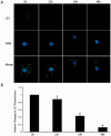
Human conditionally immortalized podocytes were put under mechanical stress. (A) Immunostaining for LC3 puncta and 4′,6-diamidino-2-phenylindole (DAPI)—binding to DNA and presenting nucleus was performed at 0 h, 12 h, 24 h, and 48 h and visualised under confocal microscope (magnification ×1200). LC3 dots are most prominent in the perinuclear and cytoplasmic regions (white arrows in merged images). LC3 puncta staining is lower at 12 h, 24 h, and 48 h, suggesting lower autophagy under mechanical stress. (B) Quantification of LC3 puncta staining showing statistically significant reduction at 24 h and 48 h (b) and statistically non-significant reduction at 12 h (a) [96]. »Reproduced with permission from Li et al., Spironolactone promotes autophagy via inhibiting PI3K/AKT/mTOR signalling pathway and reduce adhesive capacity damage in podocytes under mechanical stress. Published by Bioscience Reports—Portland Press, 2016«. The article is licensed under an open access Creative Commons CC BY 4.0 license [96].
Figure 6
Figure 6
Kidney biopsies of IgA nephropathy patients were obtained, and human mesangial cells (HMC) and human PTEC of IgA nephropathy patients were cultured, and the expression of MRs, 11β hydroxysteroid dehydrogenase (11β-HSD2), and CYP11B2 were determined using qPCR and immunofluorescence. (A) Both cell types expressed MR mRNA, whereas HMC also expressed 11β-HSD2 and CYP11B2. (B) Immunofluorescence of MR expression on PTEC (arrows) under magnification ×200. (C) Detection of all three markers in kidney biopsies of IgA nephropathy patients and controls under magnification ×200—MRs was expressed in glomeruli and tubules, whereas the other two markers were only in glomeruli. (D) A five-point scale was used to show increased glomerular CYP11B2 staining in IgA nephropathy patients compared to controls. (E) A five-point scale was used to show increased tubular MR staining in IgA nephropathy patients compared to controls. [110]. »Reproduced with permission from Leung et al., Oxidative damages in tubular epithelial cells in IgA nephropathy: role of crosstalk between angiotensin II and aldosterone; published by Journal of Translational Medicine—Springer Link, 2011«. The article is licensed under an open access Creative Commons CC BY 4.0 license [110].
Figure 7
Figure 7
Both tested MRAs can delay MR nuclear translocation. (A) After culturing human kidney GFP-MR cells for 48 h, incubation with aldosterone, spironolactone, and finerenone followed. Anti-GFP antibodies were used for immunocytochemistry with automated ArrayScan VTI fluorescent microscope (left side). DAPI was used for marking the nucleus (right side)—magnification ×20. With aldosterone treatment, complete accumulation of GFP-hMR in the nucleus was seen, but not with finerenone and spironolacotne, where it was partially located in the nucleus (B) Translocation index (average nuclear intensity/average cytoplasmic intensity) was calculated. (C) Average nuclear fluorescence intensity values calculated. V—vehicle; A—aldosterone; S—spironolactone; F—finerenone; Aldo—aldosterone; Spiro—spironolactone; Fine—finerenone [124]. »Reproduced with permission from Amazit et al., Finerenone impedes aldosterone-dependent nuclear import of the mineralocorticoid receptor and prevents genomic recruitment of steroid receptor coactivator-1; published by Journal of Biological Chemistry—currently published by Elsevier; originally published by American Society for Biochemistry and Molecular Biology., 2015«. The article is licensed under an open access Creative Commons CC BY 4.0 license [124].

References

    1. Connell J.M., Davies E. The new biology of aldosterone. J. Endocrinol. 2005;186:1–20. doi: 10.1677/joe.1.06017. - DOI - PubMed
    1. Booth R.E., Johnson J.P., Stockand J.D. Aldosterone. Adv. Physiol. Educ. 2002;26:8–20. doi: 10.1152/advan.00051.2001. - DOI - PubMed
    1. Delyani J.A. Mineralocorticoid receptor antagonists: The evolution of utility and pharmacology. Kidney Int. 2000;57:1408–1411. doi: 10.1046/j.1523-1755.2000.00983.x. - DOI - PubMed
    1. Taves M.D., Gomez-Sanchez C.E., Soma K.K. Extra-adrenal glucocorticoids and mineralocorticoids: Evidence for local synthesis, regulation, and function. Am. J. Physiol. Endocrinol. Metab. 2011;301:E11–E24. doi: 10.1152/ajpendo.00100.2011. - DOI - PMC - PubMed
    1. Brown M.J. Direct renin inhibition—A new way of targeting the renin system. J. Renin-Angiotensin-Aldosterone Syst. 2006;7:S7–S11. doi: 10.3317/jraas.2006.035. - DOI

MeSH terms

LinkOut - more resources