. 2024 Aug 22;25(16):9102.
doi: 10.3390/ijms25169102.

Development of Fusion-Based Assay as a Drug Screening Platform for Nipah Virus Utilizing Baculovirus Expression Vector System

Affiliations

Development of Fusion-Based Assay as a Drug Screening Platform for Nipah Virus Utilizing Baculovirus Expression Vector System

Indah Permata Sari et al. Int J Mol Sci. .

Abstract

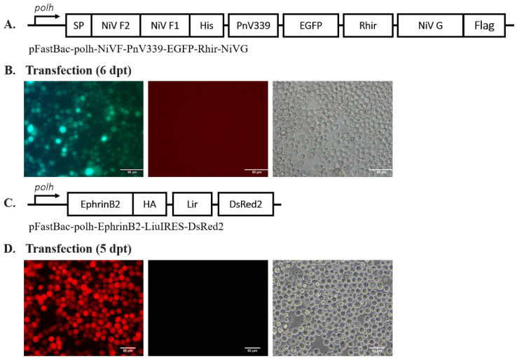
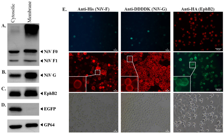
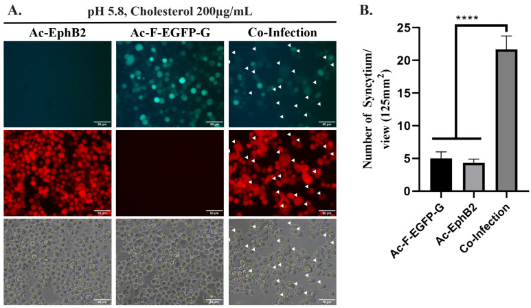
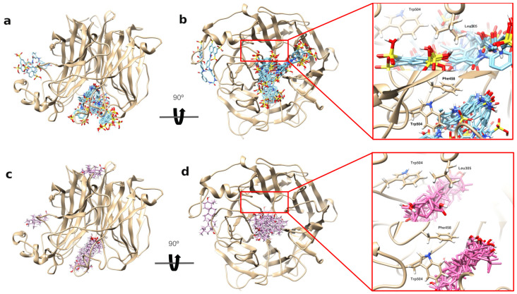
Nipah virus (NiV) is known to be a highly pathogenic zoonotic virus, which is included in the World Health Organization Research & Development Blueprint list of priority diseases with up to 70% mortality rate. Due to its high pathogenicity and outbreak potency, a therapeutic countermeasure against NiV is urgently needed. As NiV needs to be handled within a Biological Safety Level (BSL) 4 facility, we had developed a safe drug screening platform utilizing a baculovirus expression vector system (BEVS) based on a NiV-induced syncytium formation that could be handled within a BSL-1 facility. To reconstruct the NiV-induced syncytium formation in BEVS, two baculoviruses were generated to express recombinant proteins that are responsible for inducing the syncytium formation, including one baculovirus exhibiting co-expressed NiV fusion protein (NiV-F) and NiV attachment glycoprotein (NiV-G) and another exhibiting human EphrinB2 protein. Interestingly, syncytium formation was observed in infected insect cells when the medium was modified to have a lower pH level and supplemented with cholesterol. Fusion inhibitory properties of several compounds, such as phytochemicals and a polysulfonated naphthylamine compound, were evaluated using this platform. Among these compounds, suramin showed the highest fusion inhibitory activity against NiV-induced syncytium in the baculovirus expression system. Moreover, our in silico results provide a molecular-level glimpse of suramin's interaction with NiV-G's central hole and EphrinB2's G-H loop, which could be the possible reason for its fusion inhibitory activity.

Keywords: MMGBSA; Nipah virus; baculovirus expression vector system; docking; fusion inhibitor; suramin; syncytium.


Conflict of interest statement

The authors declare no conflicts of interest.

Figures

Figure 1
Figure 1
Generation of recombinant baculovirus Ac-F-EGFP-G and Ac-EphB2. (A) Schematic map of pFastBac-polh-NiVF-PnV339-EGFP-Rhir-NiVG. (B) Sf21 cells transfected with Ac-polh-NiVF-PnV339-EGFP-Rhir-NiVG bacmid were observed using the fluorescein isothiocyanate (FITC) filter at 6 dpt. Scale bar = 80 µm. (C) Schematic map of pFastBac-polh-EphrinB2-Lir-DsRed2. (D) Sf21 cells transfected with Ac-polh-EphrinB2-Lir-DsRed2 bacmid were observed using a rhodamine filter at 5 dpt. Scale bar = 50 µm.
Figure 2
Figure 2
Determination of NiV-F, NiV-G, and EphrinB2 surface expression on the infected Sf21 cell membrane. (A) Western blot result of the cytosolic and membrane protein sample collected from Ac-F-EGFP-G-infected Sf21 cells against anti-His antibody (for detecting NiV-F), and (B) anti-DDDDK antibody (for detecting NiV-G). (C) Western blot result of the cytosolic and membrane protein sample collected from Ac-EphB2-infected Sf21 cells against anti-HA antibody (for detecting EphrinB2). (D) Western blot result of the cytosolic and membrane protein sample collected from Ac-EGFP-infected Sf21 cells against anti-GFP antibody. GP64 was used as an internal control and was run as a separate gel. The SDS-PAGE was carried out using 10% acrylamide gel for NiV-F and NiV-G, and 12% acrylamide gel for EphrinB2 and EGFP. (E) IFA results for anti-His, anti-DDDDK, and anti-HA antibodies. Scale bar = 10 µm (anti-His, anti-DDDDK), 50 µm (anti-HA).
Figure 3
Figure 3
(A) Observation of NiV-induced syncytium in Ac-F-EGFP-G- and/or Ac-EphB2-infected Sf21 cells cultured in TNM-FH medium pH 5.8 and 200 µg/mL of cholesterol-supplemented TNM-FH at 27 °C for 4 dpi. Scale bar = 60 µm. White arrows indicate syncytium formation. (B) Quantification of syncytium formation in Ac-F-EGFP-G- and/or Ac-EphB2-infected Sf21 cells. Syncytium formations were quantified using three different views of the same well. Statistics were performed using one-way ANOVA, **** p-value of <0.0001. The data were presented as means with standard deviation.
Figure 4
Figure 4
Syncytium inhibitor assay. (A) Screening of fusion inhibitor activity of OA, baicalein, baicalin, and suramin. (B) Fusion inhibitory effect of different concentrations of suramin. The statistics were performed using one-way ANOVA, * p-value of <0.05, *** p-value of <0.001, **** p-value of <0.0001. The data were presented as means with standard deviation. (C) Western blot result of protein sample collected from Ac-F-EGFP-G- and Ac-EphB2-co-infected Sf21 cells against anti-His antibody (for detecting NiV-F), (D) anti-DDDDK antibody (for detecting NiV-G), and (E) anti-HA antibody (for detecting EphrinB2). GAPDH was used as the internal control and was run separately.
Figure 5
Figure 5
Top 10 docking poses of suramin and OA on NiV-G protein. (a,b) Top 10 docking poses of suramin on NiV-G. (c,d) Top 10 docking poses of OA on NiV-G. Most of the top 10 docking poses of suramin and oleanolic acid are clustered inside the central hole of NiV-G. The insets on the rightmost panels highlight key interactions with three critical residues (Leu305, Phe458, and Trp504) in NiV-G’s central hole, which are known to bind EphrinB2. Suramin and OA were shown interacting with these residues, providing further insight into their binding with NiV-G.
Figure 6
Figure 6
Top 10 docking poses of suramin and OA on human EphrinB2 and its contact distribution profile. (a,c) The top 10 docking poses of suramin closely cluster around EphrinB2’s G-H loop (residues 107–125, highlighted in red-orange in panel (a,c), in contrast to OA’s top 10 docking poses (panel c). This observation is further substantiated by the contact probability distribution illustrated in panel (b), revealing that the top 10 docking poses of suramin primarily engage with the G-H loop. The residues corresponding to the G-H loop are enclosed within an orange rectangle, highlighting a significant interaction. This stands in contrast to the contact probability distribution for OA’s poses depicted in panel (d). This contact probability distribution per residue is defined as the ratio of the number of heavy atoms of a specific EphrinB2 residue contacted by the ligand (i.e., suramin and OA) and the total number of heavy atoms contacted by the ligand for all the residues in EphrinB2. Further details on calculating this profile can be found in the Section 3.6.

Similar articles

References

    1. Negrete O.A., Levroney E.L., Aguilar H.C., Bertolotti-Ciarlet A., Nazarian R., Tajyar S., Lee B. EphrinB2 is the entry receptor for Nipah virus, an emergent deadly paramyxovirus. Nature. 2005;436:401–405. doi: 10.1038/nature03838. - DOI - PubMed
    1. Liu Q., Stone J.A., Bradel-Tretheway B., Dabundo J., Montano J.A.B., Santos-Montanez J., Biering S.B., Nicola A.V., Iorio R.M., Lu X. Unraveling a three-step spatiotemporal mechanism of triggering of receptor-induced Nipah virus fusion and cell entry. PLoS Pathog. 2013;9:e1003770. doi: 10.1371/journal.ppat.1003770. - DOI - PMC - PubMed
    1. Aguilar H.C., Iorio R.M. Henipavirus. Springer; Berlin/Heidelberg, Germany: 2012. Henipavirus membrane fusion and viral entry; pp. 79–94. - PubMed
    1. Glennon E.E., Restif O., Sbarbaro S.R., Garnier R., Cunningham A.A., Suu-Ire R.D., Osei-Amponsah R., Wood J.L., Peel A.J. Domesticated animals as hosts of henipaviruses and filoviruses: A systematic review. Vet. J. 2018;233:25–34. doi: 10.1016/j.tvjl.2017.12.024. - DOI - PubMed
    1. Chua K., Bellini W., Rota P., Harcourt B., Tamin A., Lam S., Ksiazek T., Rollin P., Zaki S., Shieh W.-J. Nipah virus: A recently emergent deadly paramyxovirus. Science. 2000;288:1432–1435. doi: 10.1126/science.288.5470.1432. - DOI - PubMed

MeSH terms

LinkOut - more resources