. 2024 Aug 22;25(16):9122.
doi: 10.3390/ijms25169122.

Automatic Detection of the EEG Spike-Wave Patterns in Epilepsy: Evaluation of the Effects of Transcranial Current Stimulation Therapy

Affiliations

Automatic Detection of the EEG Spike-Wave Patterns in Epilepsy: Evaluation of the Effects of Transcranial Current Stimulation Therapy

Elzbieta Olejarczyk et al. Int J Mol Sci. .

Abstract

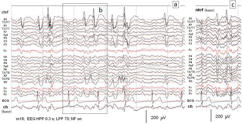
This study aims to develop a detection method based on morphological features of spike-wave (SW) patterns in the EEG of epilepsy patients and evaluate the effect of cathodal transcranial direct current stimulation (ctDCS) treatment. The proposed method is based on several simple features describing the shape of SW patterns and their synchronous occurrence on at least two EEG channels. High sensitivity, specificity and selectivity values were achieved for each patient and condition. ctDCS resulted in a significant reduction in the number of detected patterns, a decrease in spike duration and amplitude, and an increased spike mobility. The proposed method allows efficient identification of SW patterns regardless of brain condition, although the recruitment of patterns may be modified by ctDCS. This method can be useful in the clinical evaluation of ctDCS effects.

Keywords: cathodal transcranial direct current stimulation (ctDCS); electroencephalography (EEG); epilepsy; epileptiform activity; morphological features of EEG patterns; spike–wave (SW) patterns.


Conflict of interest statement

The authors declare no conflicts of interest.

Figures

Figure 1
Figure 1
(a) Segment of the EEG record with characteristic spike–wave patterns received using the reference electrode located on the chin. The record (c) presents the same data as those illustrated in (b) but recorded using the reference electrode located at the neck.
Figure 2
Figure 2
A one-second segment of the EEG record is presented in Figure 1c. Two montages are compared for the same EEG fragment: the basic montage with the reference electrode at the neck (a) and the bipolar montage (b).
Figure 3
Figure 3
The plot presents the superimposition of SW patterns from all EEG channels at the position marked in Figure 2a with the number 2. The SW pattern at electrode O1 has the largest negative amplitude of −181 μV and lasts about 70 ms. The topographical map illustrates that the SW pattern with high negative amplitude at O1 is marked with blue color, while the highest positive amplitudes are marked with red color in the right hemisphere at Fp2, F8, P8, O2.
Figure 4
Figure 4
About five-second EEG segment with SW patterns indicated by expert and/or identified by the system in patient P1 in condition PRE STIM.
Figure 5
Figure 5
The number of FP and FN patterns as a result of classification with all features compared to the classification with a set without one of the features (f1–f7) in both patients: P1 in two conditions: PRE and POST stDCS, and P2.
Figure 5
Figure 5
The number of FP and FN patterns as a result of classification with all features compared to the classification with a set without one of the features (f1–f7) in both patients: P1 in two conditions: PRE and POST stDCS, and P2.
Figure 6
Figure 6
The average SW patterns in patient P1 in two conditions: PRE and POST stDCS and in patient P2. The windows in the range from −20 ms to 20 ms from the spike maximum were considered for averaging all patterns identified in every patient separately.
Figure 7
Figure 7
Example of SW pattern on two EEG channels. The spike is from A to C, while the slower wave is from C to E.

Similar articles

References

    1. Fisher R.S., van Emde Boas W., Blume W., Elger C., Genton P., Lee P., Engel J., Jr. Epileptic seizures and epilepsy: Definitions proposed by the International League Against Epilepsy (ILAE) and the International Bureau for Epilepsy (IBE) Epilepsia. 2005;46:470–472. doi: 10.1111/j.0013-9580.2005.66104.x. - DOI - PubMed
    1. Fisher R.S., Scharfman H.E., DeCurtis M. How can we identify ictal and interictal abnormal activity? Adv. Exp. Med. Biol. 2014;813:3–23. - PMC - PubMed
    1. Kane N., Acharya J., Benickzy S., Caboclo L., Finnigan S., Kaplan P.W., Shibasaki H., Pressler R., van Putten M.J.M.A. A revised glossary of terms most commonly used by clinical electroencephalographers and updated proposal for the report format of the EEG findings. Clin. Neurophysiol. Pract. 2017;2:170–185. doi: 10.1016/j.cnp.2017.07.002. - DOI - PMC - PubMed
    1. Gotman J., Gloor P. Automatic recognition and quantification of interictal epileptic activity in the human scalp EEG. Electroenceph Clin. Neurophysiol. 1976;41:513–529. doi: 10.1016/0013-4694(76)90063-8. - DOI - PubMed
    1. Gotman J., Ives J.R., Gloor P. Automatic recognition of inter-ictal epileptic activity in prolonged EEG recordings. Electroenceph Clin. Neurophysiol. 1979;46:510–520. doi: 10.1016/0013-4694(79)90004-X. - DOI - PubMed