Review
. 2024 Jul 29;14(8):798.
doi: 10.3390/jpm14080798.

New Biomarkers in Liver Fibrosis: A Pass through the Quicksand?

Affiliations
Review

New Biomarkers in Liver Fibrosis: A Pass through the Quicksand?

Marzia Tagliaferro et al. J Pers Med. .

Abstract

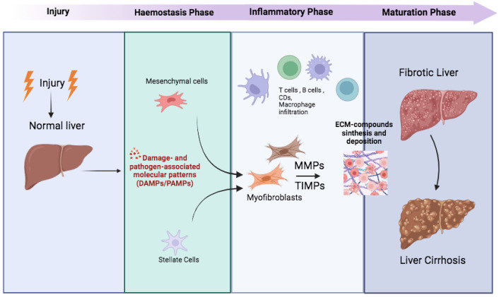
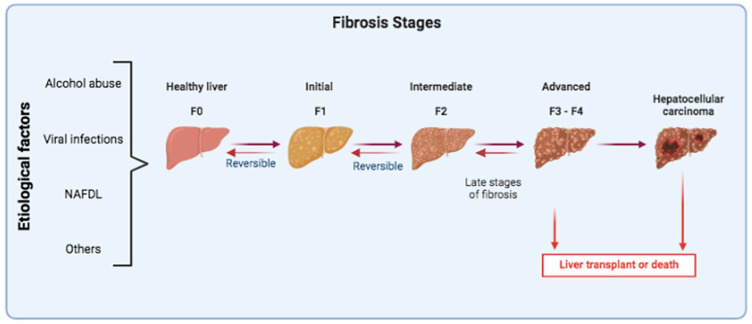
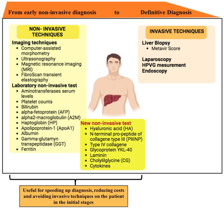
Chronic liver diseases (CLD) stem from various causes and lead to a gradual progression that ultimately may result in fibrosis and eventually cirrhosis. This process is typically prolonged and asymptomatic, characterized by the complex interplay among various cell types, signaling pathways, extracellular matrix components, and immune responses. With the prevalence of CLD increasing, diagnoses are often delayed, which leads to poor prognoses and in some cases, the need for liver transplants. Consequently, there is an urgent need for the development of novel, non-invasive methods for the diagnosis and monitoring of CLD. In this context, serum biomarkers-safer, repeatable, and more acceptable alternatives to tissue biopsies-are attracting significant research interest, although their clinical implementation is not yet widespread. This review summarizes the latest advancements in serum biomarkers for detecting hepatic fibrogenesis and advocates for concerted efforts to consolidate current knowledge, thereby providing patients with early, effective, and accessible diagnoses that facilitate personalized therapeutic strategies.

Keywords: biomarkers; extracellular matrix components; liver fibrosis.


Conflict of interest statement

The authors declare no conflicts of interest.

Figures

Figure 1
Figure 1
Cascade of fibrotic process activation in liver diseases. Physiologically, there are four distinct phases, injury, hemostasis, inflammatory phase, and maturation phase, which lead to the regeneration of damaged tissues following an injury. In the liver, when the wound response becomes pathogenic, the generation of fibrotic tissue replaces liver tissue and impairs organ function. The onset of fibrosis occurs with the activation of quiescent HSCs, i.e., resident mesenchymal cells. These cells differentiate into myofibroblasts and begin to secrete ECM constituents, particularly increasing the expression of fibrotic collagens (i.e., types III, IV, and V), fibronectins, and hyaluronic acid. Other pro-fibrotic components have also been implicated as endogenous DAMPs recognized by PRRs (pattern recognition receptors). MMPs, expressed by a variety of immune and non-immune cells, degrade ECM components, including collagen and fibronectin, making them essential for tissue remodeling. The balance between MMPs and TIMPs in the liver plays a vital role in the induction of liver fibrosis.
Figure 2
Figure 2
Liver fibrosis stages. Persistent liver damage, regardless of its various etiologies, leads to a progressive deposition of fibrous tissue and alteration of the normal liver parenchyma. The METAVIR fibrosis score classifies fibrosis into five possible stages: F0 = no fibrosis, F1 = portal fibrosis without septa, F2 = portal fibrosis with rare septa, F3 = numerous septa without cirrhosis, and F4 = cirrhosis. Ultimately, cirrhosis can progress to liver cancer. The progression of liver fibrosis can be interrupted or reversed with the elimination of the hit in the early stages F1 and F2, while the advanced stages are not easily reversible, in fact only proper therapies could revert the stage of disease. The only alternative for the patient’s recovery is a liver transplant.
Figure 3
Figure 3
Summary of techniques used for staging liver fibrosis. In the left panel, non-invasive techniques such as imaging techniques and laboratory tests are mentioned. The new laboratory tests useful for staging are mentioned in the dark yellow box. Invasive techniques are listed in the right panel.

References

    1. Iredale J.P. Models of liver fibrosis: Exploring the dynamic nature of inflammation and repair in a solid organ. J. Clin. Investig. 2007;117:539–548. doi: 10.1172/JCI30542. - DOI - PMC - PubMed
    1. Novo E., Cannito S., Paternostro C., Bocca C., Miglietta A., Parola M. Cellular and molecular mechanisms in liver fibrogenesis. Arch. Biochem. Biophys. 2014;548:20–37. doi: 10.1016/j.abb.2014.02.015. - DOI - PubMed
    1. Bocca C., Novo E., Miglietta A., Parola M. Angiogenesis and Fibrogenesis in Chronic Liver Diseases. Cell Mol. Gastroenterol. Hepatol. 2015;1:477–488. doi: 10.1016/j.jcmgh.2015.06.011. - DOI - PMC - PubMed
    1. Vallonthaiel A.G., Baloda V., Singh L., Yadav R., Kilambi R., Battu S., Sreenivas V., Pal S., Acharya S.K., DattaGupta S., et al. Histological analyses of trucut liver biopsies from patients with noncirrhotic portal fibrosis and extra-hepatic portal vein obstruction. Indian J. Pathol. Microbiol. 2021;64((Suppl. 1)):S12–S135. doi: 10.4103/IJPM.IJPM_387_20. - DOI - PubMed
    1. Pocino K., Stefanile A., Basile V., Napodano C., D’Ambrosio F., Di Santo R., Callà C.A.M., Gulli F., Saporito R., Ciasca G., et al. Cytokines and Hepatocellular Carcinoma: Biomarkers of a Deadly Embrace. J. Pers. Med. 2023;13:5. doi: 10.3390/jpm13010005. - DOI - PMC - PubMed

LinkOut - more resources