. 2024 Aug 9;14(8):847.
doi: 10.3390/jpm14080847.

Starvation and Inflammation Modulate Adipose Mesenchymal Stromal Cells' Molecular Signature

Affiliations

Starvation and Inflammation Modulate Adipose Mesenchymal Stromal Cells' Molecular Signature

Simona Piccolo et al. J Pers Med. .

Abstract

Mesenchymal stromal cells (MSCs) and their released factors (secretome) are intriguing options for regenerative medicine approaches based on the management of inflammation and tissue restoration, as in joint disorders like osteoarthritis (OA). Production strategy may modulate cells and secretome fingerprints, and for the latter, the effect of serum removal by starvation used in clinical-grade protocols has been underestimated. In this work, the effect of starvation on the molecular profile of interleukin 1 beta (IL1β)-primed adipose-derived MSCs (ASCs) was tested by assessing the expression level of 84 genes related to secreted factors and 84 genes involved in defining stemness potential. After validation at the protein level, the effect of starvation modulation in the secretomes was tested in a model of OA chondrocytes. IL1β priming in vitro led to an increase in inflammatory mediators' release and reduced anti-inflammatory potential on chondrocytes, features reversed by subsequent starvation. Therefore, when applying serum removal-based clinical-grade protocols for ASCs' secretome production, the effects of starvation must be carefully considered and investigated.

Keywords: adipose mesenchymal stromal cells; chondrocytes; inflammation; osteoarthritis; secretome; starvation.


Conflict of interest statement

The authors declare no conflicts of interest. The funders had no role in the design of the study; in the collection, analyses, or interpretation of data; in the writing of the manuscript; or in the decision to publish the results.

Figures
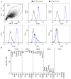
Figure 1
Figure 1
ASCs’ immunophenotype: (A) ASCs were positive for MSC markers CD44, CD73, CD90, and CD105, and tissue-resident/early-passage ASC marker CD34. Plots illustrate the results from a representative donor. (B) No difference was observed under the analyzed conditions (N = 3).
Figure 2
Figure 2
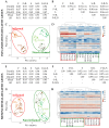
Correlation between samples and conditions: (A) Intra-group and (B) inter-group correlation analyses for the expression of inflammation-related genes; N = 3, mean ± SD. “r” Pearson values are shown. (C) PCA performed on normalized Ct values for inflammation genes. The X and Y axes show principal component 1 and principal component 2, which explain 47.3% and 15.3% of the total variance. (D) Hierarchical clustering performed on normalized Ct values for inflammation genes. Higher Ct means lower amount, and lower Ct means higher amount. (E) Intra-group and (F) inter-group correlation analyses for the expression of mesenchymal stem cell-related genes; N = 3, mean ± SD. “r” Pearson values are shown. (G) PCA performed on normalized Ct values for mesenchymal genes. The X and Y axes show principal component 1 and principal component 2, which explain 35.8% and 13.0% of the total variance. (H) Hierarchical clustering performed on normalized Ct values for mesenchymal genes. Higher Ct means lower amount, and lower Ct means higher amount.
Figure 3
Figure 3
Correlation between samples and conditions for significantly modulated genes: (A) PCA performed on ln(FC + 1) values, with FC calculated vs. condition F. The X and Y axes show principal component 1 and principal component 2, which explain 87.7% and 9.7% of the total variance. (B) Hierarchical clustering performed on ln(FC + 1) values, with FC calculated vs. condition F.
Figure 4
Figure 4
Quantitative analysis of factors modulated by IL1β and reversed by subsequent starvation. CCL2, CXCL2, IL6, IL8, CSF2, and CSF3 levels detected as pg/mL were measured by ELISA assays. In the absence of plots, the proteins were undetectable or below the lower limit of detection of the assay. Under ANOVA analysis, significance was set for p-value ≤ 0.05 (§ for p-value ≤ 0.1, * ≤ 0.05, ** ≤ 0.01, *** ≤ 0.001 and **** ≤ 0.0001. N = 3).
Figure 5
Figure 5
Correlation between conditions for significantly modulated genes in inflamed chondrocytes treated with secretomes: (A) Inter-group correlation analysis of the modulation of OA-related genes in chondrocytes treated with IL1β alone or with IL1β and secretomes with respect to untreated (CTRL) cells. “r” Pearson values are shown. (B) PCA performed on ln(FC + 1) values, with FC calculated vs. condition CTRL. The X and Y axes show principal component 1 and principal component 2, which explain 91.6% and 6.6% of the total variance, respectively. (C) Hierarchical clustering performed on ln(FC + 1) values, with FC calculated vs. condition CTRL. The scale bar’s maximum for ln(FC + 1) values was set to 2.5.

References

    1. Merimi M., El-Majzoub R., Lagneaux L., Moussa Agha D., Bouhtit F., Meuleman N., Fahmi H., Lewalle P., Fayyad-Kazan M., Najar M. The Therapeutic Potential of Mesenchymal Stromal Cells for Regenerative Medicine: Current Knowledge and Future Understandings. Front. Cell Dev. Biol. 2021;9:661532. doi: 10.3389/fcell.2021.661532. - DOI - PMC - PubMed
    1. Han Y., Yang J., Fang J., Zhou Y., Candi E., Wang J., Hua D., Shao C., Shi Y. The secretion profile of mesenchymal stem cells and potential applications in treating human diseases. Signal Transduct. Target. Ther. 2022;7:92. doi: 10.1038/s41392-022-00932-0. - DOI - PMC - PubMed
    1. Murphy M.B., Moncivais K., Caplan A.I. Mesenchymal stem cells: Environmentally responsive therapeutics for regenerative medicine. Exp. Mol. Med. 2013;45:54. doi: 10.1038/emm.2013.94. - DOI - PMC - PubMed
    1. Miceli V., Zito G., Bulati M., Gallo A., Busà R., Iannolo G., Conaldi P.G. Different priming strategies improve distinct therapeutic capabilities of mesenchymal stromal/stem cells: Potential implications for their clinical use. World J. Stem Cells. 2023;15:400–420. doi: 10.4252/wjsc.v15.i5.400. - DOI - PMC - PubMed
    1. Sanz-Nogués C., O’Brien T. Current good manufacturing practice considerations for mesenchymal stromal cells as therapeutic agents. Biomater. Biosyst. 2021;2:100018. doi: 10.1016/j.bbiosy.2021.100018. - DOI - PMC - PubMed

LinkOut - more resources