. 2024 Aug 8;14(16):1725.
doi: 10.3390/diagnostics14161725.

Anatomical Relationships of the Proximal Attachment of the Hamstring Muscles with Neighboring Structures: From Ultrasound, Anatomical and Histological Findings to Clinical Implications

Affiliations

Anatomical Relationships of the Proximal Attachment of the Hamstring Muscles with Neighboring Structures: From Ultrasound, Anatomical and Histological Findings to Clinical Implications

Maribel Miguel-Pérez et al. Diagnostics (Basel). .

Abstract

Background: Injuries of the proximal attachment of the hamstring muscles are common. The present study aimed to investigate the relationship of the proximal attachment of the hamstring muscles with neighboring structures comprehensively.

Methods: A total of 97 hemipelvis from 66 cryopreserved specimens were evaluated via ultrasound, anatomical and histological samples.

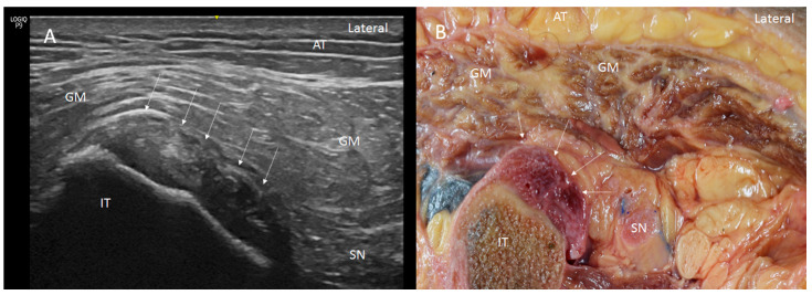
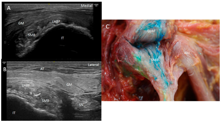
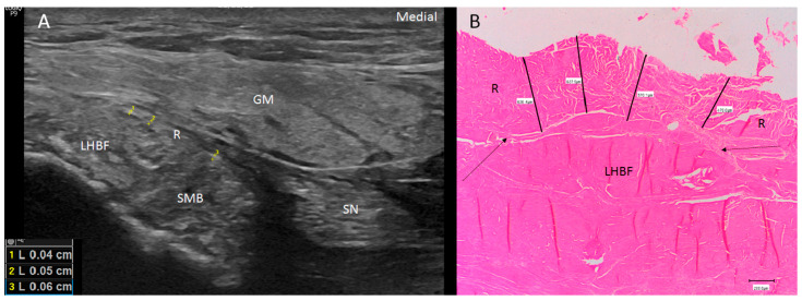
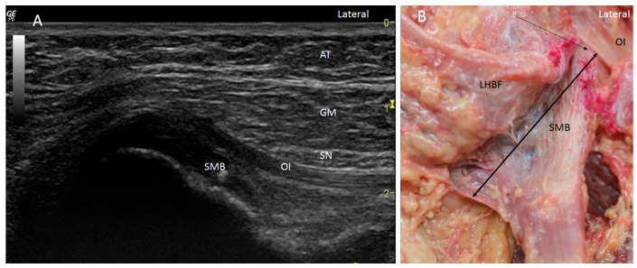
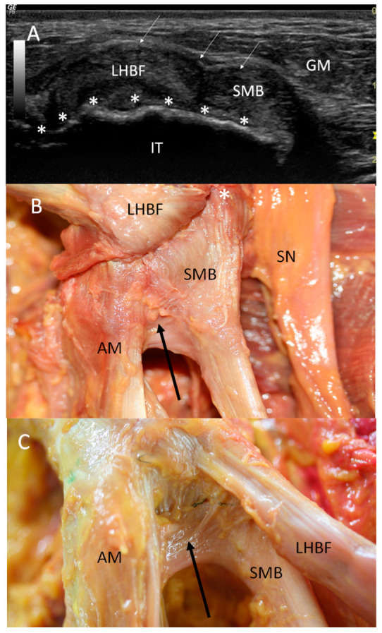
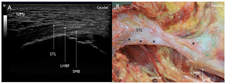
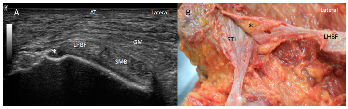
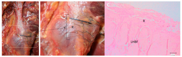
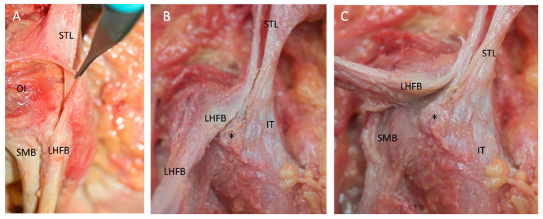
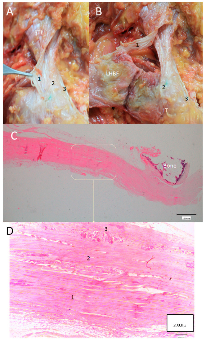
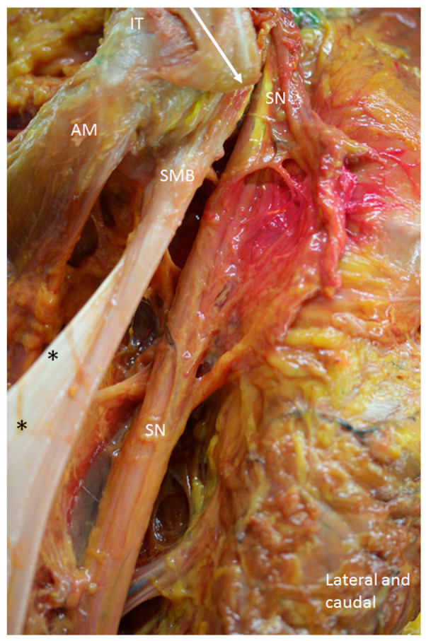
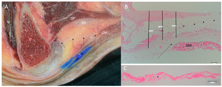
Results: The proximal attachment of the hamstring muscles presents a hyperechogenic line surrounding the origin of the semimembranosus and the long head of the biceps femoris muscles, as well as another hyperechogenic line covering the sciatic nerve. The anatomical and histological study confirms the ultrasound results and shows different layers forming the sacrotuberous ligament. Furthermore, it shows that the proximal attachment of the semimembranosus muscle has a more proximal origin than the rest of the hamstring muscles. Moreover, this muscle shares fibers with the long head of the biceps femoris muscle and expands to the adductor magnus muscle. The histological analysis also shows the dense connective tissue of the retinaculum covering the long head of the biceps femoris and semimembranosus muscles, as well as the expansion covering the sciatic nerve.

Conclusions: These anatomical relationships could explain injuries at the origin of the hamstring muscles.

Keywords: adductor magnus; biceps femoris; hamstring; ischial tuberosity; musculoskeletal; sacrotuberous ligament; sciatic nerve; tendinopathy; ultrasound.


Conflict of interest statement

The authors declare no conflicts of interest.

Figures

Figure 1
Figure 1
(A) Ultrasound view in short axis of the right tendon of long head of biceps femoris (LHBF) and semimembranosus (SMB) muscles seen as two round hyperechogenic structures similar to a mask or the shape of a goat’s leg at the ischial tuberosity (IT). They are surrounded and isolated by a hyperechogenic line that fixed them to the bone (white arrows). Adipose tissue (AT), gluteus maximus muscle (GM), sciatic nerve (SN), (B) Anatomical section of the hip showing the LHBF and SMB tendons fixed by the retinaculum (white arrows).
Figure 2
Figure 2
(A,B) Two ultrasound examples in the short axis of the tendons of the long head of biceps femoris (LHBF) and the semimembranosus (SMB) muscles, presenting an irregular origin at the ischial tuberosity (IT) with several hyperechogenic structures that were compatible with a degeneration of the tendon: (A) left side; (B) right side. (C) Dissection of the left area showing, after moving the LHBF, a very irregular insertion of the tendon of the SMB with all the blue dye widespread. AM: adductor maximus muscle.
Figure 3
Figure 3
(A) Ultrasound view of the retinaculum (R) as a hyperechogenic line that fixed them to the bone (as well as the different points where it was measured (yellow numbers 1, 2 and 3). Tendon of long head of biceps femoris (LHBF) and semimembranosus muscle (SMB). Right side: gluteus maximus muscle (GM). (B) Histological view of the retinaculum and its measures of 636.4 µ 637.9 µ, 570.1 µ and 470 µ. It is composed of dense connective tissue, and it is possible to observe the perpendicular direction of its fibers with the fibers of the tendon of LHBF and their close relation (black arrows).
Figure 4
Figure 4
(A) The expansion over the sciatic nerve (SN) on the left side. An ultrasound study allows us to visualize this expansion (yellow =), which goes from the origin of the hamstrings (H) (yellow arrow) to the gluteus maximus muscle (GM) (white arrow). IT: Ischial tuberosity. (B) Anatomical study allowing us to see this expansion (black arrows) over the sciatic nerve on the left side. It is possible to visualize that it goes from the origin of the hamstrings (H) (yellow arrow) to the gluteus maximus muscle (GM) (white arrow).
Figure 5
Figure 5
(A) Ultrasound study in the short axis on the right side showing the high origin semimembranosus muscle (SMB). It goes up even as far as to be placed under the obturator internus muscle (OI). SN: sciatic nerve GM: gluteus maximus muscle; AT: adipose tissue. (B) Anatomical view of the long and high origin of the tendon of the SMB (black arrow) under the OI after moving the LHBF medially on the right side.
Figure 6
Figure 6
(A) Ultrasound view in the short axis showing the high origin of the right tendon of the semimembranosus muscle (SMB) (white *) at the ischial tuberosity (IT). LHBF: the tendon of the long head of biceps femoris; white arrow: retinaculum (white arrow); GM: gluteus maximus muscle. (B) Anatomic view of the origin after moving up the LHBF; it is possible to visualize the high origin of the SMB (white *) and the deep connections (black arrow) with the origin of the adductor maximus muscle (AM) (laterally, the sciatic nerve). (C) Anatomic view to observe the deep relation between LHBF and the SMB and the SMB and AM (black arrow). Both anatomical views are on the right side.
Figure 7
Figure 7
(A) Ultrasound view at the long axis showing that the left sacrotuberous ligament (STL) is seen as a hyperechogenic structure that continues caudally with the tendon of the long head biceps femoris (LHBF). Also, the tendon of the semimembranosus muscle is located as a hyperechogenic structure under LHBFB. GlMy: Gluteus maximus muscle (B) Anatomical dissection showing that the fibers of the left STL cross between them with a lambda (λ)-shaped bifurcation. Then, the superficial and medial side of the STL (white arrow) goes on caudally with the LHBF. Moreover, it is possible to observe that the lateral and the deeper part of the sacrotuberous ligament inserts at the ischial tuberosity (black *), showing an x shape. The deeper layers of the ligament (black *) insert at the ischial tuberosity (IT).
Figure 8
Figure 8
(A) Ultrasound view of the sacrotuberous ligament insertion at the vertex (white *) of the ischial tuberosity (IT) in the short axis tendon of the semimembranosus muscle (SMB) on the right side (gluteus maximus muscle). AT: Adipose tissue. (B) Anatomical dissection of the sacrotuberous ligament on the right side showing the possibility to separate different layers of the STL. The most superficial layer of the STL (black *) follows with the LHBF, and it is separated from the deep layer with adipose tissue (black *) and other layers of the STL inserted at the vertex of the IT (white *).
Figure 9
Figure 9
(A) Anatomical view of the retinaculum (R) at the ischial tuberosity (IT) on the right side. (B) It is possible to see the retinaculum increasing with the transversal fibers fixing the origin of the LHBF and SMB (in this case, it is possible to see a transversal black line corresponding to an injected vessel). (C) Histological study showing the retinaculum as dense connective tissue in a very close relation with the LHBF.
Figure 10
Figure 10
(A) The accurate dissection of the STL showing that the most superficial layer of the sacrotuberous ligament (STL) is continuous with the long tendon of biceps femoris (LHBF) superficially on the right side. OI: Obturator internus. (B) STL inserted at the LHBF as and also has a insertion as a cord in the vertex (black *) of the ischial tuberosity and at the medial side of the ischial tuberosity (IT). (C) After moving medially the LHBF it is possible to observe all the origin extension of the tendon of semimembranosus muscle (SMB).
Figure 11
Figure 11
(A,B) The anatomical study showing different layers (1, 2, 3) of the sacrotuberous ligament (STL) that insert at different points on the right side. They have to be separated with a scalpel; however, it is very evident in the difference between layers 1 and 2. Layer 1 follows with the superficial tendinous fibers of the tendon of the long head biceps femoris (LHBF). Layer 2 inserts at the vertex of the ischial tuberosity and layer 3 at the medial side of the ischial tuberosity. (C) Microscopical study showing the dense connective tissue of the STL, seen transversally, and its relationship with the sacrum bone. (D) A higher vision of the STL allowing us to see the three layers separated, in this case, by dense, less-organize connective tissue.
Figure 12
Figure 12
Anatomical view of the posterior and right side of the hip. The gluteus maximus muscle, the long head of the biceps femoris and the semitendinosus has been removed to observe the tendon of the semimembranosus muscle (SMB). It has a rounder shape at the more superior and lateral part (white arrow), which distally became thinner, and a thin membrane medially (black *) that originates at the lateral side of the ischial tuberosity (IT). AM: Adductor maximus muscles. The sciatic nerve (SN) at the lateral side has a close relationship with the SMB.
Figure 13
Figure 13
The expansion over the sciatic nerve (SN) (black arrows) was observed and measured: (A) anatomical view of the expansion going from the origin of the hamstrings (H) to the gluteus maximus muscle (GM) on the right side; (B) histological study allowing us to identify several thin and irregular layers of the dense connective tissue separated by adipose tissue (black *) (200.0 µ). (C) histological view of the expansion with similar morphology (2.000 mm).

Similar articles

Cited by

References

    1. Williams M.A., Naffaa L. Ischial Tuberosity Avulsion Fracture Mimicking Calcified Mass on Plain Films: A Case Report. Cureus. 2024;16:e53165. doi: 10.7759/cureus.53165. - DOI - PMC - PubMed
    1. Maniar N., Carmichael D.S., Hickey J.T., Timmins R.G., San Jose A.J., Dickson J., Opar D. Incidence and prevalence of hamstring injuries in field-based team sports: A systematic review and meta-analysis of 5952 injuries from over 7 million exposure hours. Br. J. Sports Med. 2023;57:109–116. doi: 10.1136/bjsports-2021-104936. - DOI - PubMed
    1. Sheean A.J., Arner J.W., Bradley J.P. Proximal Hamstring Tendon Injuries: Diagnosis and Management. Arthroscopy. 2021;37:435–437. doi: 10.1016/j.arthro.2020.12.201. - DOI - PubMed
    1. Fletcher A.N., Cheah J.W., Nho S.J., Mather R.C. Proximal Hamstring Injuries. Clin. Sports Med. 2021;40:339–361. doi: 10.1016/j.csm.2021.01.003. - DOI - PubMed
    1. Ekstrand J., Hägglund M., Waldén M. Epidemiology of muscle injuries in professional football (soccer) Am. J. Sports Med. 2011;39:1226–1232. doi: 10.1177/0363546510395879. - DOI - PubMed