Reliability of Kaiser Score in Assessing Additional Breast Lesions Identified on Staging MRI in Patients with Breast Cancer
- PMID: 39202214
- PMCID: PMC11353333
- DOI: 10.3390/diagnostics14161726
Reliability of Kaiser Score in Assessing Additional Breast Lesions Identified on Staging MRI in Patients with Breast Cancer
Abstract
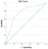
(1) Background: The Kaiser score is a user-friendly tool that evaluates lesions on breast MRI and has been studied in the general population and a few specific clinical scenarios. We aim to evaluate the performance of the Kaiser score in the characterization of additional lesions identified on staging breast MRI. (2) Methods: The Kaiser score of the biopsied additional lesions identified on staging MRI in recently diagnosed breast cancer patients was retrospectively determined. Statistical analysis was performed to evaluate the diagnostic capability of the Kaiser score and whether it is affected by different imaging and pathological parameters of the additional and the index lesion. (3) Results: Seventy-six patients with ninety-two MRI-detected lesions constitute the studied population. There was a statistically significant difference in the Kaiser score between benign and malignant lesions, irrespective of the pathology of the index cancer (p = 0.221) or the size and the imaging features of the additional lesion. Using a cutoff of 5 and above for suspicious lesions, biopsy could have been avoided in 34/92 lesions. (4) Conclusions: The Kaiser score can assist radiologists in the evaluation of additional MRI lesions identified in recently diagnosed breast cancer patients, thus decreasing the number of unneeded biopsies and delays in definitive surgical management.
Keywords: Kaiser score; Tree flowchart; additional cancer; breast cancer; staging breast MRI.
Conflict of interest statement
The authors declare no conflicts of interest.
Figures


Similar articles
-
The Kaiser score reliably excludes malignancy in benign contrast-enhancing lesions classified as BI-RADS 4 on breast MRI high-risk screening exams.Eur Radiol. 2020 Nov;30(11):6052-6061. doi: 10.1007/s00330-020-06945-z. Epub 2020 Jun 6. Eur Radiol. 2020. PMID: 32504098 Free PMC article.
-
Application of the Kaiser score by MRI in patients with breast lesions by ultrasound and mammography.Diagn Interv Radiol. 2022 Jul;28(4):322-328. doi: 10.5152/dir.2022.201075. Diagn Interv Radiol. 2022. PMID: 35950276 Free PMC article.
-
Impact of the Kaiser score on clinical decision-making in BI-RADS 4 mammographic calcifications examined with breast MRI.Eur Radiol. 2020 Mar;30(3):1451-1459. doi: 10.1007/s00330-019-06444-w. Epub 2019 Dec 3. Eur Radiol. 2020. PMID: 31797077 Free PMC article.
-
Multiparametric MRI in detection and staging of prostate cancer.Dan Med J. 2017 Feb;64(2):B5327. Dan Med J. 2017. PMID: 28157066 Review.
-
How to use the Kaiser score as a clinical decision rule for diagnosis in multiparametric breast MRI: a pictorial essay.Insights Imaging. 2018 Jun;9(3):325-335. doi: 10.1007/s13244-018-0611-8. Epub 2018 Apr 3. Insights Imaging. 2018. PMID: 29616496 Free PMC article. Review.
References
-
- Breast Cancer. [(accessed on 26 June 2024)]. Available online: https://www.who.int/news-room/fact-sheets/detail/breast-cancer.
LinkOut - more resources
Full Text Sources

