Review
. 2024 Aug 16;14(16):1786.
doi: 10.3390/diagnostics14161786.

Computed Tomography of Contemporary Occupational Lung Disease: A Pictorial Review

Affiliations
Review

Computed Tomography of Contemporary Occupational Lung Disease: A Pictorial Review

Jimin Lee et al. Diagnostics (Basel). .

Abstract

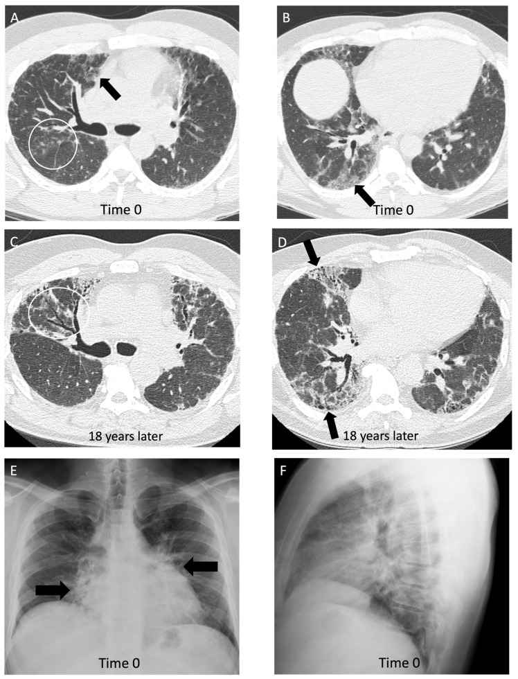
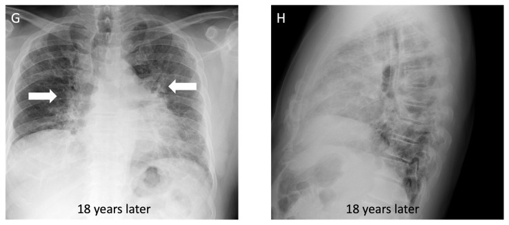
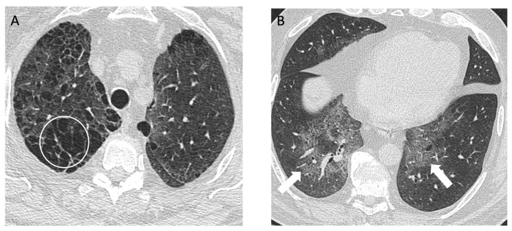
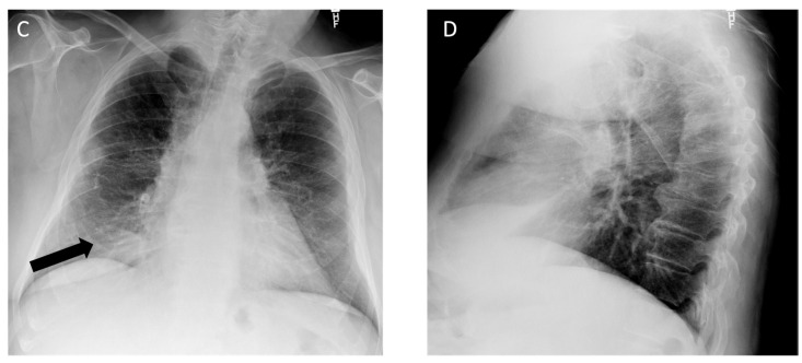
Occupational lung disease remains one of the most common work-related illnesses and accounts for most deaths from occupational illness. Occupational lung diseases often have delayed manifestation over decades and nonspecific clinical presentations, making it challenging for clinicians to promptly identify the disease and implement preventive measures. Radiologists play a crucial role in identifying and diagnosing occupational lung diseases, allowing for removal of the exposure and early medical intervention. In this review, we share our clinical and radiologic approach to diagnosing occupational lung disease and its subtypes. A collection of sample cases of occupational lung diseases commonly encountered in the modern era at a large Canadian university hospital is included to facilitate understanding. This review will provide radiologists with valuable insights into recognizing and diagnosing occupational lung diseases.

Keywords: asbestosis; hypersensitivity pneumonitis; occupational lung disease; pneumoconiosis; silicosis; work-related asthma.


Conflict of interest statement

M.M. reports consulting fees from AstraZeneca and honoraria from Boehringer Ingelheim, unrelated to the current work. All other authors declare no relevant relationships.

Figures

Figure 1
Figure 1
A 45-year-old non-smoking male, previously a stone mason and countertop stone cutter, diagnosed with complicated silicosis and progressive massive fibrosis. (A) Axial computed tomography image shows conglomerate bilateral upper zone masses with associated architectural distortion consistent with progressive massive fibrosis (white arrows); (B) coronal image demonstrates the upper lung zone predominance; (C) chest radiograph demonstrates profuse small circumscribed pulmonary nodules with an upper lung zone predominance.
Figure 2
Figure 2
A 73-year-old male with a history of remote 16 pack year smoking history and asbestos exposure as a retired plumber working on pipe and broiler insulation, diagnosed with asbestos-related pleural disease. (A) Axial computed tomography shows asbestos plaques on the right (long arrows) and diffuse pleural thickening on the left (short arrows) on the soft tissue window; (B) on the lung window, a subpleural band opacity is seen in the left lower lobe (black arrow); (C) posteroanterior view and (D) lateral view chest radiographs show diffuse pleural thickening with calcification, best appreciated by costophrenic angle blunting (black arrows).
Figure 3
Figure 3
An 83-year-old male with 75 pack year smoking history and asbestos exposure from construction work, diagnosed with asbestosis. (A) Axial computed tomography image demonstrates subpleural reticulation (black arrows) and mild traction bronchiectasis (white circle) of right lung in keeping with mild asbestosis; (B) mediastinal windows demonstrate a calcified pleural plaque along the right diaphragm (white arrow); (C) posteroanterior and (D) lateral chest radiograph demonstrate mild reticular markings in the basal lung consistent with mild fibrosis, best appreciated in the left lateral costophrenic angle (white circle).
Figure 4
Figure 4
An 89-year-old non-smoking male with asbestos exposure from construction work, diagnosed with epithelioid mesothelioma, later confirmed on video-assisted thoracoscopic surgery biopsy. (A) On axial computed tomography, there is a partly calcified pleural plaque anteriorly on the right (black arrow) and volume loss in the left lung (asterisks) related to a rind of pleural thickening; (B) on the left, the rind of pleural thickening (arrow) has a nodular appearance consistent with mesothelioma and there is a small pleural effusion after thoracentesis that drained 1.2 L of fluid; follow-up (C) posteroanterior and (D) lateral view radiographs one week later demonstrate a reaccumulated moderate-to-large size pleural effusion opacifying the left mid-to-lower lung zone (asterisks).
Figure 5
Figure 5
A 43-year-old non-smoking male with 15-year history of tungsten carbide exposure as a metal grinding tool maker, diagnosed with hard metal lung disease, which was confirmed with surgical lung biopsy. (A,B) Axial computed tomography images demonstrate patchy ground glass opacities (arrows) and small centrilobular nodules (white circle) with minimal architectural distortion and traction bronchiectasis; (C,D) computed tomography images 18 years later demonstrate long-term evolution of fibrosis, with development of traction bronchiectasis (white circle) and mild honeycombing (black arrows) on the background of ground glass opacity and coarse reticulation; (E) posteroanterior view and (F) lateral view chest radiographs show a perihilar and lower lung zone predominant reticular abnormality (black arrows); (G) posteroanterior view and (H) lateral view chest radiographs 18 years later demonstrate a reduction in lung volumes, and the prior interstitial opacities appear coarser and more prominent, particularly in the perihilar lung (white arrows).
Figure 5
Figure 5
A 43-year-old non-smoking male with 15-year history of tungsten carbide exposure as a metal grinding tool maker, diagnosed with hard metal lung disease, which was confirmed with surgical lung biopsy. (A,B) Axial computed tomography images demonstrate patchy ground glass opacities (arrows) and small centrilobular nodules (white circle) with minimal architectural distortion and traction bronchiectasis; (C,D) computed tomography images 18 years later demonstrate long-term evolution of fibrosis, with development of traction bronchiectasis (white circle) and mild honeycombing (black arrows) on the background of ground glass opacity and coarse reticulation; (E) posteroanterior view and (F) lateral view chest radiographs show a perihilar and lower lung zone predominant reticular abnormality (black arrows); (G) posteroanterior view and (H) lateral view chest radiographs 18 years later demonstrate a reduction in lung volumes, and the prior interstitial opacities appear coarser and more prominent, particularly in the perihilar lung (white arrows).
Figure 6
Figure 6
A 56-year-old male with a history of grinding aluminum for 5 years diagnosed with desquamative interstitial pneumonia (DIP) on surgical lung biopsy. There was a 10 pack year smoking history, but it was remote to the presentation, and the DIP was attributed to occupational exposure. (A) Axial computed tomography of the upper lung zone reveals emphysema (white circle), and the (B) basal lung zone demonstrates extensive ground glass opacity (arrows) with subtle small cysts or lucencies on close inspection; (C) posteroanterior and (D) lateral view chest radiographs 6 months later show persistent basilar opacities (black arrow).
Figure 6
Figure 6
A 56-year-old male with a history of grinding aluminum for 5 years diagnosed with desquamative interstitial pneumonia (DIP) on surgical lung biopsy. There was a 10 pack year smoking history, but it was remote to the presentation, and the DIP was attributed to occupational exposure. (A) Axial computed tomography of the upper lung zone reveals emphysema (white circle), and the (B) basal lung zone demonstrates extensive ground glass opacity (arrows) with subtle small cysts or lucencies on close inspection; (C) posteroanterior and (D) lateral view chest radiographs 6 months later show persistent basilar opacities (black arrow).
Figure 7
Figure 7
A 56-year-old male with a remote 20 pack year smoking history and a history of beryllium exposure who was working with electronics, diagnosed with chronic beryllium disease. Axial computed tomography shows interstitial fibrosis characterized by (A) peripheral subpleural and (B) peribronchovascular distribution of reticulation and mild traction bronchiectasis (arrows) with a basal predominance.
Figure 8
Figure 8
A 62-year-old non-smoking male with metal vapor exposure in the workplace was diagnosed with non-fibrotic hypersensitivity pneumonitis on surgical lung biopsy. (A) CT of lung bases demonstrates diffuse ground glass abnormality posteriorly (white circle); (B) follow up CT 6 months after being removed from the workplace demonstrates compete resolution of the opacities; (C) chest radiograph shows a subtle increase in lung density at the bases (asterisks) with relatively preserved lung volumes.
Figure 9
Figure 9
A 76-year-old non-smoking female was diagnosed with farmer’s lung, proven with biopsy. Computed tomography of the upper lobe demonstrates (A) peribronchovascular reticulations (white circle) with mosaic lung attenuation and (B) mild diffuse subpleural reticulations (black arrows); (C) posteroanterior and (D) lateral view radiographs demonstrate diffusely coarse reticulations and fibrosis (asterisks) in the upper lung zone with some mild volume loss. A staple line in left lower lobe is noted from a previous lung biopsy for diagnosis of farmer’s lung disease (black arrow).

Similar articles

References

    1. Blanc P.D., Annesi-Maesano I., Balmes J.R., Cummings K.J., Fishwick D., Miedinger D., Murgia N., Naidoo R.N., Reynolds C.J., Sigsgaard T., et al. The Occupational Burden of Nonmalignant Respiratory Diseases. An Official American Thoracic Society and European Respiratory Society Statement. Am. J. Respir. Crit. Care Med. 2019;199:1312–1334. doi: 10.1164/rccm.201904-0717ST. - DOI - PMC - PubMed
    1. Matyga A.W., Chelala L., Chung J.H. Occupational Lung Diseases: Spectrum of Common Imaging Manifestations. Korean J. Radiol. 2023;24:795–806. doi: 10.3348/kjr.2023.0274. - DOI - PMC - PubMed
    1. Handra C.-M., Gurzu I.-L., Chirila M., Ghita I. Silicosis: New Challenges from an Old Inflammatory and Fibrotic Disease. Front. Biosci. 2023;28:96. doi: 10.31083/j.fbl2805096. - DOI - PubMed
    1. Hoy R.F., Jeebhay M.F., Cavalin C., Chen W., Cohen R.A., Fireman E., Go L.H.T., León-Jiménez A., Menéndez-Navarro A., Ribeiro M., et al. Current global perspectives on silicosis—Convergence of old and newly emergent hazards. Respirology. 2022;27:387–398. doi: 10.1111/resp.14242. - DOI - PMC - PubMed
    1. Walkoff L., Hobbs S. Chest Imaging in the Diagnosis of Occupational Lung Diseases. Clin. Chest Med. 2020;41:581–603. doi: 10.1016/j.ccm.2020.08.007. - DOI - PubMed

LinkOut - more resources