Review
. 2024 Aug 16;14(16):1788.
doi: 10.3390/diagnostics14161788.

Extraosseous Plasmacytomas: A Radiologist's Perspective-A Narrative Review of the Literature

Affiliations
Review

Extraosseous Plasmacytomas: A Radiologist's Perspective-A Narrative Review of the Literature

Konstantinos Stefanidis et al. Diagnostics (Basel). .

Abstract

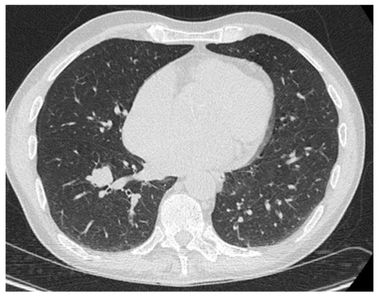
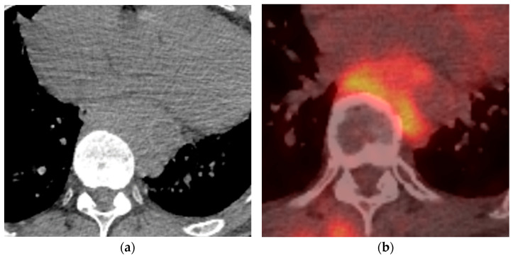
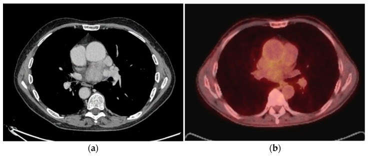
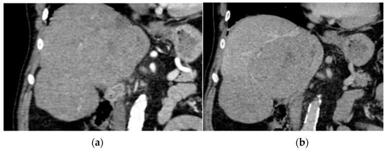
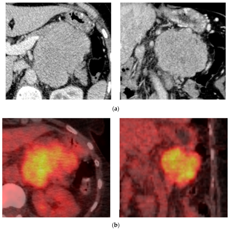
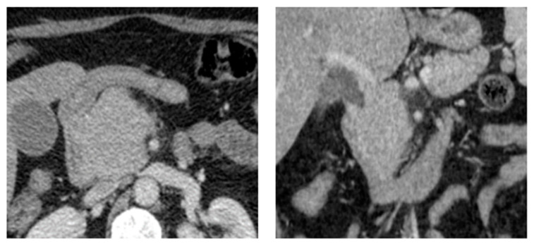
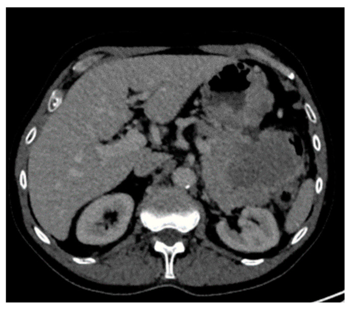
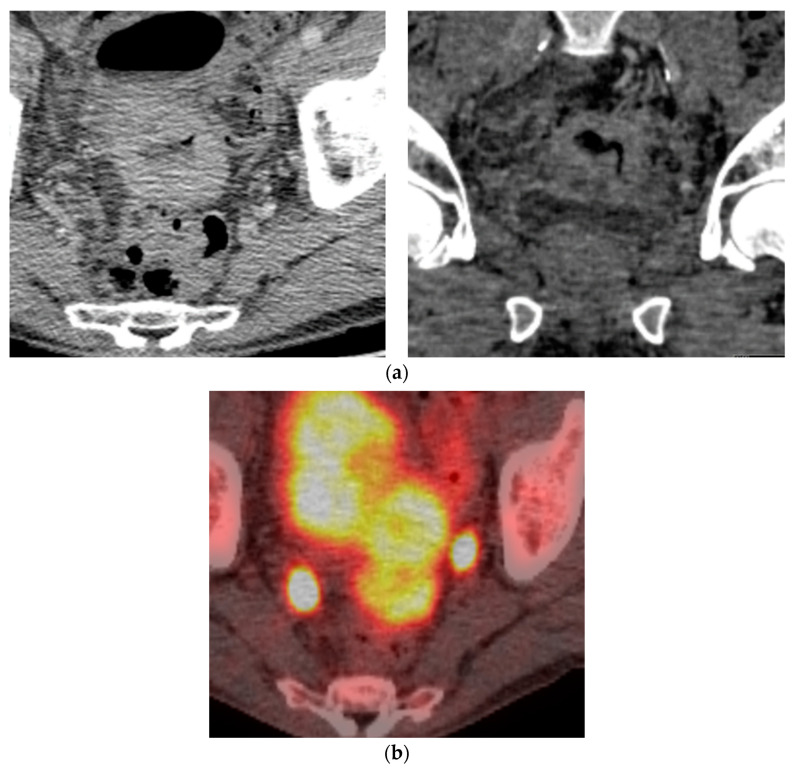
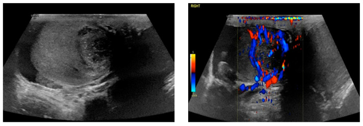
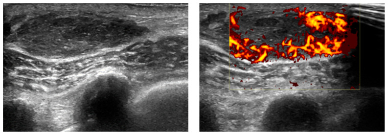
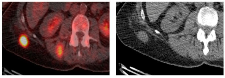
Extraosseous plasmacytomas (EPs) are rare neoplasms originating from plasma cells, often associated with multiple myeloma. EPs are classified into three subtypes: extramedullary myeloma, solitary extramedullary plasmacytoma (SEP), and multiple solitary plasmacytomas. They can manifest in various anatomical sites, including the lung, mediastinum, breast, liver, pancreas, stomach, mesentery, kidney, small and large bowel, testis, and soft tissue. Despite their rarity, EPs present a diagnostic challenge due to their non-specific imaging appearances, which can mimic other neoplastic and inflammatory conditions. This review aims to describe the radiographic features of EPs in the chest, abdomen, and pelvis based on a thorough analysis of the existing literature. While imaging plays a crucial role in the detection and characterization of EPs, histological confirmation is necessary to differentiate them from other neoplastic entities. The review underscores the importance of considering EPs in the differential diagnosis, particularly in patients with a history of multiple myeloma. Understanding the imaging characteristics of EPs is essential for accurate diagnosis and appropriate management. Early imaging is crucial in these patients to exclude the possibility of EP, as timely diagnosis can significantly impact patient outcomes.

Keywords: extraosseous plasmacytomas; multiple myeloma; radiographic features.


Conflict of interest statement

The authors declare no conflicts of interest.

Figures

Figure 1
Figure 1
Biopsy-proven lung EP, case from our institution. Axial CT image demonstrates a lobulated lung nodule in the right lower lobe.
Figure 2
Figure 2
Biopsy-proven Mediastinal EP, case from our institution. (a) Unenhanced axial CT shows a posterior mediastinal soft tissue mass; (b) Axial PET-CT image shows heterogenous increased FDG-uptake in the posterior mediastinal mass.
Figure 3
Figure 3
Biopsy-proven Mediastinal EP, case from our institution (a) Axial CT image shows an enhancing soft tissue lesion in the subcarinal space; (b) PET-CT image at the same level shows the mediastinal mass with mild heterogenous avidity.
Figure 4
Figure 4
Biopsy-proven Liver EP, case from our institution (a) Arterial phase coronal CT shows two well-defined liver lesions, both of which show mild enhancement; (b) Portal venous phase coronal CT shows a very mild washout of contrast in both lesions.
Figure 5
Figure 5
Biopsy-proven pancreas EP, case from our institution. (a) Post-contrast axial and coronal CT shows a well-defined homogenous lobular mass at the body of the pancreas; (b) Axial and coronal PET-CT shows increased FDG uptake in the pancreatic lesion.
Figure 6
Figure 6
Biopsy-proven Pancreas EP, case from our institution. Axial and coronal post-contrast CT shows a well-defined homogenous mass at the pancreatic head. The coronal imaging demonstrates associated dilatation of the common bile duct.
Figure 7
Figure 7
Biopsy-proven Gastric EP, case from our institution (Axial contrast-enhanced CT image shows a heterogeneous enhancing gastric mass with exophytic and intraluminal components.
Figure 8
Figure 8
Biopsy-proven Colon EP, case from our institution. (a) Axial and coronal post-contrast CT demonstrates homogenous mural thickening of the sigmoid colon with resultant luminal narrowing; (b) Axial PET CT shows avid uptake in the sigmoid colon lesion.
Figure 9
Figure 9
Biopsy-proven Testicular EP, case from our institution. Testicular ultrasound with B-mode and color Doppler shows a heterogeneous, hypoechoic lesion with punctuate calcifications and peripheral vascularity.
Figure 10
Figure 10
Biopsy-roven Soft tissue EP, case from our institution. Soft tissue ultrasound with B-mode and color Doppler shows a well-defined, heterogeneous, ovoid lesion with increased peripheral vascularity.
Figure 11
Figure 11
Biopsy-proven Soft tissue EP, case from our institution. Axial non-contrast CT and PET-CT show a well-defined, PET-avid soft tissue nodular lesion in the subcutaneous tissue.

Similar articles

Cited by

References

    1. Sevcikova S., Minarik J., Stork M., Jelinek T., Pour L., Hajek R. Extramedullary disease in multiple myeloma—Controversies and future directions. Blood Rev. 2019;36:32–39. doi: 10.1016/j.blre.2019.04.002. - DOI - PubMed
    1. Weinstock M., Ghobrial I.M. Extramedullary multiple myeloma. Leuk. Lymphoma. 2013;54:1135–1141. doi: 10.3109/10428194.2012.740562. - DOI - PubMed
    1. Touzeau C., Moreau P. How I treat extramedullary myeloma. Blood. 2016;127:971–976. doi: 10.1182/blood-2015-07-635383. - DOI - PubMed
    1. International Myeloma Working G. Criteria for the classification of monoclonal gammopathies, multiple myeloma and related disorders: A report of the International Myeloma Working Group. Br. J. Haematol. 2003;121:749–757. doi: 10.1046/j.1365-2141.2003.04355.x. - DOI - PubMed
    1. Gozdziuk K., Kedra M., Rybojad P., Sagan D. A rare case of solitary plasmacytoma mimicking a primary lung tumor. Ann. Thorac. Surg. 2009;87:e25–e26. doi: 10.1016/j.athoracsur.2008.11.066. - DOI - PubMed

LinkOut - more resources