Three-Dimensional Morphometric Analysis of the Volar Cortical Shape of the Lunate Facet of the Distal Radius
- PMID: 39202290
- PMCID: PMC11353463
- DOI: 10.3390/diagnostics14161802
Three-Dimensional Morphometric Analysis of the Volar Cortical Shape of the Lunate Facet of the Distal Radius
Abstract
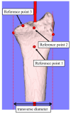
In cases of distal radius fractures, the fixation of the volar lunate facet fragment is crucial for preventing volar subluxation of the carpal bones. This study aims to clarify the sex differences in the volar morphology of the lunate facet of the distal radius and its relationship with the transverse diameter of the distal radius. Sixty-four CT scans of healthy wrists (30 males and 34 females) were evaluated. Three-dimensional (3D) images of the distal radius were reconstructed from the CT data. We defined reference point 1 as the starting point of the inclination toward the distal volar edge, reference point 2 as the volar edge of the joint on the bone axis, and reference point 3 as the volar edge of the distal radius lunate facet. From the 3D coordinates of reference points 1 to 3, the bone axis distance, volar-dorsal distance, radial-ulnar distance, 3D straight-line distance, and inclination angle were measured. The transverse diameter of the radius was measured, and its correlations with the parameters were evaluated. It was found that in males, compared to females, the transverse diameter of the radius is larger and the protrusion of the volar lunate facet is greater. This suggests that the inclination of the volar surface is steeper in males and that the volar locking plate may not fit properly with the volar cortical bone of the lunate facet, necessitating additional fixation.
Keywords: computer analysis; distal radius fracture; lunate facet; volar locking plate.
Conflict of interest statement
No benefits in any form have been received or will be received from a commercial party related directly or indirectly to the subject of this article.
Figures


Similar articles
-
Volar Lunate Facet Fractures of the Distal Radius: Fracture Mapping Using 3D CT Scans.J Wrist Surg. 2022 Jan 20;11(6):484-492. doi: 10.1055/s-0041-1742228. eCollection 2022 Dec. J Wrist Surg. 2022. PMID: 36504531 Free PMC article.
-
Clinical Outcomes in Distal Radius Fractures Accompanied by Volar Lunate Facet Fragments: A Comparison between Dorsal and Volar Displaced Fractures.J Hand Surg Asian Pac Vol. 2020 Dec;25(4):417-422. doi: 10.1142/S2424835520500447. J Hand Surg Asian Pac Vol. 2020. PMID: 33115368
-
Is There a Critical Dorsal Lunate Facet Size in Distal Radius Fractures That Leads to Dorsal Carpal Subluxation? A Biomechanical Study of the Dorsal Critical Corner.J Hand Surg Am. 2025 Feb;50(2):240.e1-240.e6. doi: 10.1016/j.jhsa.2023.07.001. Epub 2023 Aug 16. J Hand Surg Am. 2025. PMID: 37589617
-
Volar-Ulnar Approach for Fixation of the Volar Lunate Facet Fragment in Distal Radius Fractures: A Technical Tip.J Hand Surg Am. 2016 Dec;41(12):e491-e500. doi: 10.1016/j.jhsa.2016.09.007. J Hand Surg Am. 2016. PMID: 27916152 Review.
-
A Loop-Wiring Technique for Volarly Displaced Distal Radius Fractures With Small Thin Volar Marginal Fragments.J Hand Surg Am. 2020 Mar;45(3):261.e1-261.e7. doi: 10.1016/j.jhsa.2019.10.022. Epub 2019 Dec 16. J Hand Surg Am. 2020. PMID: 31859052 Review.
Cited by
-
A novel approach for three-dimensional evaluation of reduction morphology in distal radius fracture.BMC Musculoskelet Disord. 2025 Feb 15;26(1):156. doi: 10.1186/s12891-025-08367-8. BMC Musculoskelet Disord. 2025. PMID: 39955499 Free PMC article.
-
A Novel Method to Represent the Three-Dimensional Inclination of the Distal Radius Joint Surface.Diagnostics (Basel). 2025 Feb 1;15(3):345. doi: 10.3390/diagnostics15030345. Diagnostics (Basel). 2025. PMID: 39941275 Free PMC article.
References
Grants and funding
LinkOut - more resources
Full Text Sources
Medical

