Review
. 2024 Aug 21;14(16):1821.
doi: 10.3390/diagnostics14161821.

Line-Field Confocal Optical Coherence Tomography: A New Skin Imaging Technique Reproducing a "Virtual Biopsy" with Evolving Clinical Applications in Dermatology

Affiliations
Review

Line-Field Confocal Optical Coherence Tomography: A New Skin Imaging Technique Reproducing a "Virtual Biopsy" with Evolving Clinical Applications in Dermatology

Simone Cappilli et al. Diagnostics (Basel). .

Abstract

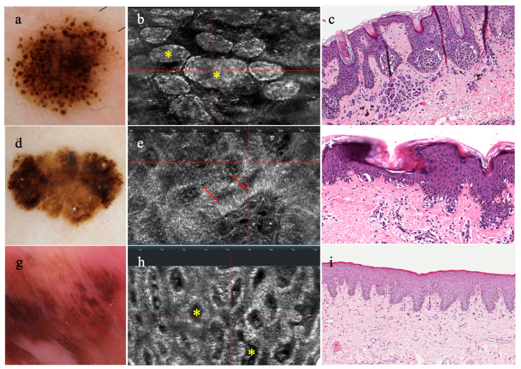
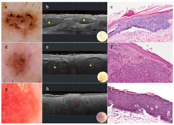
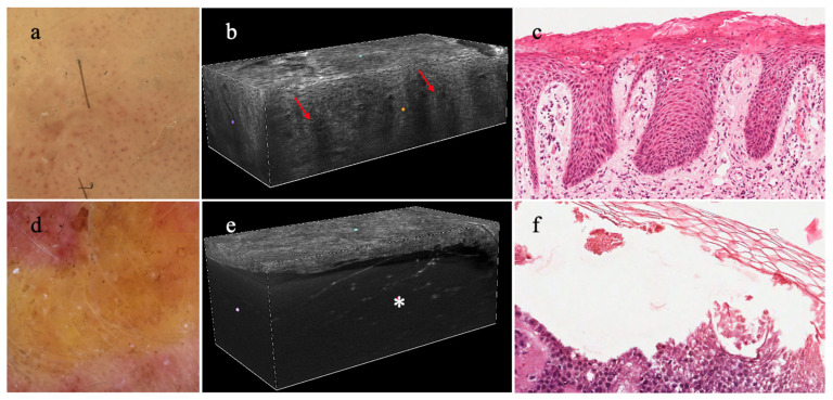
Background: Line-field confocal optical coherence tomography is a novel technology able to reproduce a "virtual biopsy" of the skin. The aim of this review is to explore the application of line-field confocal optical coherence tomography (LC-OCT) in various skin diseases, covering skin cancers, inflammatory and infectious skin diseases, genetic diseases, cosmetic procedures, and less common disorders.

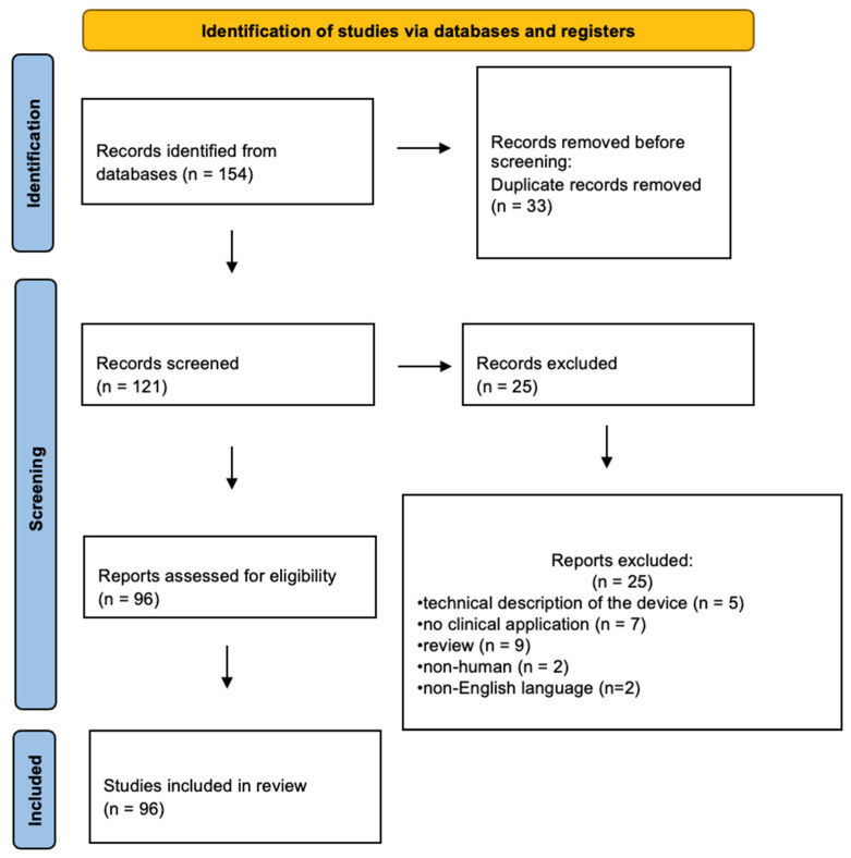
Methods: Study selection was conducted based on LC-OCT and using pertinent MeSh terms, following Preferred Reporting Items for Systematic Reviews and Meta-Analyses (PRISMA) guidelines from inception to March 2024; to evaluate the quality and risk of bias of studies, Quality Assessment of Diagnostic Accuracy Studies-2 (QUADAS-2) was used.

Results: the search retrieved 154 papers according to the selection criteria; after removing publications by one or more of the exclusion criteria, a total of 96 studies were found to be suitable for the analysis.

Conclusions: Increasing evidence supports the use of LC-OCT as an adjunctive diagnostic tool for the in vivo diagnosis of a variety of skin tumors. As this device can be considered a "bridge" between dermoscopy and histopathology, widening applications in numerous fields of clinical dermatology, including inflammatory skin disease treatment, presurgical mapping, cosmetic procedures, and monitoring of non-invasive therapies, have been explored.

Keywords: clinical dermatology; diagnostic imaging; histology of the skin; line-field confocal optical coherence tomography; review; virtual biopsy.


Conflict of interest statement

Peris received consulting fees and honoraria from Abbvie, Almirall, Biogen, Celgene, Janssen Galderma, Novartis, Lilly, Novartis, Pierre Fabre, Sandoz, Sanofi and Sun Pharma outside of the submitted work. All the remaining authors declare that they have no conflicts of interest relevant to this manuscript.

Figures

Figure 1
Figure 1
PRISMA flowchart diagram (adapted from Page et al., 2021) [20].
Figure 2
Figure 2
Dermatoscopy, horizontal LC-OCT, and histopathology of compound nevus, melanoma, genital melanosis: (a) compound nevus with globular pattern (b) associated with junctional nests (yellow asterisks) in LC-OCT and (c) histopathology; (d) melanoma showing atypical network and eccentric hyperpigmented areas in dermatoscopy, (e) due to atypical junctional melanocytes (red arrows) in LC-OCT (f) clearly seen in histopathology; (g) melanosis with dermatoscopic features of parallel pattern (h) that reveals a draped pattern (yellow asterisks) in LC-OCT, (i) corresponding to hyperpigmentation of the basal keratinocytes in histopathology.
Figure 3
Figure 3
Dermatoscopy, vertical LC-OCT, and histopathology of superficial basal cell carcinoma (sBCC), nodular basal cell carcinoma (nBCC), and Bowen disease: (a) sBCC showing leaf-like areas and erosions in dermatoscopy, (b) identified under LC-OCT as hemispheric lobules attached to the epidermis (yellow asterisks), (c) relating to tumor islands in histopathology; (d) nBCC with arborizing micro vessels and brown structureless areas in dermatoscopy, (e) visualized as ovoid-shaped lobules separated from the epidermis (yellow asterisks) in LC-OCT, and (f) histopathology; (g) Bowen disease with dermatoscopic pattern of glomerular vessels and a scaly surface, (h) revealing hyperkeratosis and dilated vessels (red arrows) in LC-OCT, and (i) histopathology.
Figure 4
Figure 4
Dermatoscopy, 3D LC-OCT, and histopathology of plaque psoriasis and bullous pemphigoid: (a) plaque psoriasis with a regular distribution of red dots and white scales in dermatoscopy, (b) showing elongation of the dermal papillae (red arrows) and dilated vessels in LC-OCT (c) clearly observed in histopathology; (d) bullous pemphigoid characterized by a yellow translucent structureless area in dermatoscopy, (e) justified by a junctional split creating a bulla with floating elements in LC-OCT (white asterisk), (f) as seen in histopathology.

Similar articles

Cited by

References

    1. Jartarkar S.R., Patil A., Wollina U., Gold M.H., Stege H., Grabbe S., Goldust M. New diagnostic and imaging technologies in dermatology. J. Cosmet. Dermatol. 2021;20:3782–3787. doi: 10.1111/jocd.14499. - DOI - PubMed
    1. Schneider S.L., Kohli I., Hamzavi I.H., Council M.L., Rossi A.M., Ozog D.M. Emerging imaging technologies in dermatology: Part I: Basic principles. J. Am. Acad. Dermatol. 2019;80:1114–1120. doi: 10.1016/j.jaad.2018.11.042. - DOI - PMC - PubMed
    1. Zavattaro E., Veronese F., Savoia P. Editorial: Non-invasive diagnostic tools in the management of skin disorders. Front. Med. 2023;10:1271195. doi: 10.3389/fmed.2023.1271195. - DOI - PMC - PubMed
    1. Greenwood J.D., Merry S.P., Boswell C.L. Skin Biopsy Techniques. Prim. Care. 2022;49:1–22. doi: 10.1016/j.pop.2021.10.001. - DOI - PubMed
    1. Elston D.M., Stratman E.J., Miller S.J. Skin biopsy: Biopsy issues in specific diseases. J. Am. Acad. Dermatol. 2016;74:1–16. doi: 10.1016/j.jaad.2015.06.033. Erratum in J. Am. Acad. Dermatol. 2016, 75, 854. - DOI - PubMed

LinkOut - more resources