

Perivascular Fat: A Novel Risk Factor for Coronary Artery Disease
- PMID: 39202318
- PMCID: PMC11353828
- DOI: 10.3390/diagnostics14161830
Perivascular Fat: A Novel Risk Factor for Coronary Artery Disease
Abstract
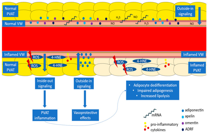
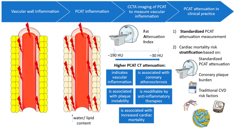
Perivascular adipose tissue (PVAT) interacts with the vascular wall and secretes bioactive factors which regulate vascular wall physiology. Vice versa, vascular wall inflammation affects the adjacent PVAT via paracrine signals, which induce cachexia-type morphological changes in perivascular fat. These changes can be quantified in pericoronary adipose tissue (PCAT), as an increase in PCAT attenuation in coronary computed tomography angiography images. Fat attenuation index (FAI), a novel imaging biomarker, measures PCAT attenuation around coronary artery segments and is associated with coronary artery disease presence, progression, and plaque instability. Beyond its diagnostic capacity, PCAT attenuation can also ameliorate cardiac risk stratification, thus representing an innovative prognostic biomarker of cardiovascular disease (CVD). However, technical, biological, and anatomical factors are weakly related to PCAT attenuation and cause variation in its measurement. Thus, to integrate FAI, a research tool, into clinical practice, a medical device has been designed to provide FAI values standardized for these factors. In this review, we discuss the interplay of PVAT with the vascular wall, the diagnostic and prognostic value of PCAT attenuation, and its integration as a CVD risk marker in clinical practice.
Keywords: atherosclerosis; cardiovascular disease; coronary computed tomography angiography; fat attenuation index; pericoronary adipose tissue attenuation; perivascular adipose tissue.
Conflict of interest statement
The authors declare no conflicts of interest.
Figures
Similar articles
-
Perivascular Adipose Tissue and Coronary Atherosclerosis: from Biology to Imaging Phenotyping.Curr Atheroscler Rep. 2019 Nov 19;21(12):47. doi: 10.1007/s11883-019-0817-3. Curr Atheroscler Rep. 2019. PMID: 31741080 Free PMC article. Review.
-
Diagnostic potential of pericoronary adipose tissue mean attenuation for coronary atherosclerotic heart disease: a comparative analysis with the fat attenuation index.Quant Imaging Med Surg. 2025 Apr 1;15(4):3148-3160. doi: 10.21037/qims-24-828. Epub 2025 Mar 28. Quant Imaging Med Surg. 2025. PMID: 40235807 Free PMC article.
-
State-of-the-art review article. Atherosclerosis affecting fat: What can we learn by imaging perivascular adipose tissue?J Cardiovasc Comput Tomogr. 2019 Sep-Oct;13(5):288-296. doi: 10.1016/j.jcct.2019.03.006. Epub 2019 Mar 29. J Cardiovasc Comput Tomogr. 2019. PMID: 30952610 Free PMC article. Review.
-
High pericoronary adipose tissue attenuation on computed tomography angiography predicts cardiovascular events in patients with type 2 diabetes mellitus: post-hoc analysis from a prospective cohort study.Cardiovasc Diabetol. 2022 Mar 18;21(1):44. doi: 10.1186/s12933-022-01478-9. Cardiovasc Diabetol. 2022. PMID: 35303857 Free PMC article.
-
Pericoronary adipose tissue attenuation assessed by dual-layer spectral detector computed tomography is a sensitive imaging marker of high-risk plaques.Quant Imaging Med Surg. 2021 May;11(5):2093-2103. doi: 10.21037/qims-20-860. Quant Imaging Med Surg. 2021. PMID: 33936990 Free PMC article.
Cited by
-
Cardiac magnetic resonance quantified epicardial fat volume is associated with complex coronary artery disease among diabetics.Cardiovasc Diabetol. 2025 Feb 7;24(1):64. doi: 10.1186/s12933-025-02606-x. Cardiovasc Diabetol. 2025. PMID: 39920759 Free PMC article.
-
The Perivascular Fat Attenuation Index: Bridging Inflammation and Cardiovascular Disease Risk.J Clin Med. 2025 Jul 4;14(13):4753. doi: 10.3390/jcm14134753. J Clin Med. 2025. PMID: 40649128 Free PMC article. Review.
-
Inflammation in atherosclerotic cardiovascular disease: From diagnosis to treatment.Eur J Clin Invest. 2025 Jul;55(7):e70020. doi: 10.1111/eci.70020. Epub 2025 Mar 8. Eur J Clin Invest. 2025. PMID: 40055964 Free PMC article. Review.
-
Coronary Inflammation and Cardiovascular Events in Patients Without Obstructive Coronary Artery Disease.Curr Cardiol Rep. 2025 Mar 7;27(1):68. doi: 10.1007/s11886-025-02221-y. Curr Cardiol Rep. 2025. PMID: 40053166 Free PMC article. Review.
-
Vascular effects of perivascular adipose tissue-derived chemerin in obesity-associated cardiovascular disease.Cardiovasc Diabetol. 2025 Jun 13;24(1):249. doi: 10.1186/s12933-025-02814-5. Cardiovasc Diabetol. 2025. PMID: 40514684 Free PMC article. Review.
References
Publication types
LinkOut - more resources
Full Text Sources
