

Whole-Exome Analysis for Polish Caucasian Patients with Retinal Dystrophies and the Creation of a Reference Genomic Database for the Polish Population
- PMID: 39202371
- PMCID: PMC11353931
- DOI: 10.3390/genes15081011
Whole-Exome Analysis for Polish Caucasian Patients with Retinal Dystrophies and the Creation of a Reference Genomic Database for the Polish Population
Abstract
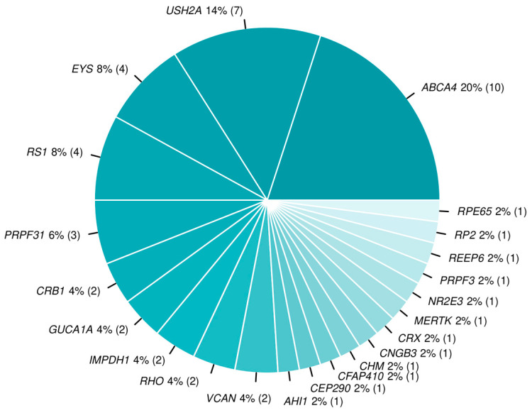
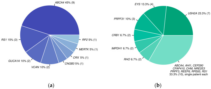
We present the results of the first study of a large cohort of patients with inherited retinal dystrophies (IRD) performed for the Polish population using whole-exome sequencing (WES) in the years 2016-2019. Moreover, to facilitate such diagnostic analyses and enable future application of gene therapy and genome editing for IRD patients, a Polish genomic reference database (POLGENOM) was created based on whole-genome sequences of healthy Polish Caucasian nonagenarians and centenarians. The newly constructed database served as a control, providing a comparison for variant frequencies in the Polish population. The diagnostic yield for the selected group of IRD patients reached 64.9%. The study uncovered the most common pathogenic variants in ABCA4 and USH2A in the European population, along with several novel causative variants. A significant frequency of the ABCA4 complex haplotype p.(Leu541Pro; Ala1038Val) was observed, as well as that of the p.Gly1961Glu variant. The first VCAN causative variant NM_004385.5:c.4004-2A>G in Poland was found and described. Moreover, one of the first patients with the RPE65 causative variants was identified, and, in consequence, could receive the dedicated gene therapy. The availability of the reference POLGENOM database enabled comprehensive variant characterisation during the NGS data analysis, confirming the utility of a population-specific genomic database for enhancing diagnostic accuracy. Study findings suggest the significance of genetic testing in elder patients with unclear aetiology of eye diseases. The combined approach of NGS and the reference genomic database can improve the diagnosis, management, and future treatment of IRDs.
Keywords: WES; WGS; inherited retinal dystrophy; population database; retinitis pigmentosa.
Conflict of interest statement
E.M., M.B.-G., K.S., E.G., P.Ł., R.S., M.J., M.Z., and A.B.-C. are employees of Genomed S.A. A.B.-C. and M.Z. keep stocks in Genomed S.A. The funders had no role in the design of the study; in the collection, analyses, or interpretation of data; in the writing of the manuscript; or in the decision to publish the results.
Figures
References
-
- RetNet The Retinal Information Network. [(accessed on 2 April 2024)]. Available online: https://web.sph.uth.edu/RetNet/
-
- Tuupanen S., Gall K., Sistonen J., Saarinen I., Kämpjärvi K., Wells K., Merkkiniemi K., von Nandelstadh P., Sarantaus L., Känsäkoski J., et al. Prevalence of RPGR-Mediated Retinal Dystrophy in an Unselected Cohort of Over 5000 Patients. Transl. Vis. Sci. Technol. 2022;11:6. doi: 10.1167/tvst.11.1.6. - DOI - PMC - PubMed
MeSH terms
Substances
Grants and funding
LinkOut - more resources
Full Text Sources
