Review
. 2024 Aug 2;15(8):1017.
doi: 10.3390/genes15081017.

A Closer Look into White Adipose Tissue Biology and the Molecular Regulation of Stem Cell Commitment and Differentiation

Affiliations
Review

A Closer Look into White Adipose Tissue Biology and the Molecular Regulation of Stem Cell Commitment and Differentiation

Presley D Dowker-Key et al. Genes (Basel). .

Abstract

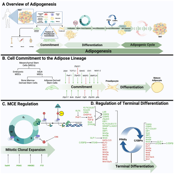
White adipose tissue (WAT) makes up about 20-25% of total body mass in healthy individuals and is crucial for regulating various metabolic processes, including energy metabolism, endocrine function, immunity, and reproduction. In adipose tissue research, "adipogenesis" is commonly used to refer to the process of adipocyte formation, spanning from stem cell commitment to the development of mature, functional adipocytes. Although, this term should encompass a wide range of processes beyond commitment and differentiation, to also include other stages of adipose tissue development such as hypertrophy, hyperplasia, angiogenesis, macrophage infiltration, polarization, etc.… collectively, referred to herein as the adipogenic cycle. The term "differentiation", conversely, should only be used to refer to the process by which committed stem cells progress through distinct phases of subsequent differentiation. Recognizing this distinction is essential for accurately interpreting research findings on the mechanisms and stages of adipose tissue development and function. In this review, we focus on the molecular regulation of white adipose tissue development, from commitment to terminal differentiation, and examine key functional aspects of WAT that are crucial for normal physiology and systemic metabolic homeostasis.

Keywords: adipogenesis; metabolism; mitotic clonal expansion; stem commitment and differentiation; white adipose tissue.


Conflict of interest statement

The authors declare no conflicts of interest.

Figures

Figure 1
Figure 1
Molecular controls and regulations of adipocyte development. (A) WAT is found throughout the human body and congregates in specific sites referred to as fat depots. The two major depots, subcutaneous (SAT) and visceral (VAT), both serve distinct metabolic and nonmetabolic roles in the body. Although WAT depots are heterogenous, adipocytes account for more than 90% of WAT volume [67], with the remaining collectively known as the stromal vascular fraction (SVF). The SVF is composed of various cell types that contribute to WAT’s growth, repair, renewal, and functioning. Importantly, a subpopulation of immature adipocytes resides in this fraction, awaiting the appropriate cues to undergo maturation. (A) In a highly strategic manner, an adipose precursor cell is (1) committed to the adipogenic lineage and then sequentially undergoes (2) proliferation, (3) clonal expansion (MCE), (4) early- and (5) late-stage differentiation. (6) Adipocyte expansion via hyperplasia and/or hypertrophy, along with processes such as cell turnover, angiogenesis and macrophage infiltration attribute to the establishment of mature adipocytes and WAT (All together referred to as the adipogenic cycle). (B,C) Various molecular regulators have been shown to either promote or inhibit stem/stromal cell commitment as well as MCE, both critical phases of adipocyte differentiation. (D) Terminal differentiation is largely characterized by PPARγ and C/EBPα, notably the master transcription factors. Equally as important is their maintenance by upstream modulators that either promote or inhibit the master transcription factors. Figure created with BioRender.com.

Similar articles

Cited by

References

    1. Azeez O.I., Meintjes R., Chamunorwa J.P. Fat body, fat pad and adipose tissues in invertebrates and vertebrates: The nexus. Lipids Health Dis. 2014;13:71. doi: 10.1186/1476-511X-13-71. - DOI - PMC - PubMed
    1. Ottaviani E., Malagoli D., Franceschi C. The evolution of the adipose tissue: A neglected enigma. Gen. Comp. Endocrinol. 2011;174:1–4. doi: 10.1016/j.ygcen.2011.06.018. - DOI - PubMed
    1. Chusyd D.E., Wang D., Huffman D.M., Nagy T.R. Relationships between Rodent White Adipose Fat Pads and Human White Adipose Fat Depots. Front. Nutr. 2016;3:10. doi: 10.3389/fnut.2016.00010. - DOI - PMC - PubMed
    1. Wronska A., Kmiec Z. Structural and biochemical characteristics of various white adipose tissue depots. Acta Physiol. 2012;205:194–208. doi: 10.1111/j.1748-1716.2012.02409.x. - DOI - PubMed
    1. Martins T., Castro-Ribeiro C., Lemos S., Ferreira T., Nascimento-Gonçalves E., Rosa E., Oliveira P.A., Antunes L.M. Murine Models of Obesity. Obesities. 2022;2:127–147. doi: 10.3390/obesities2020012. - DOI

LinkOut - more resources