. 2024 Aug 2;15(8):1019.
doi: 10.3390/genes15081019.

Tracking Ovine Pulmonary Adenocarcinoma Development Using an Experimental Jaagsiekte Sheep Retrovirus Infection Model

Affiliations

Tracking Ovine Pulmonary Adenocarcinoma Development Using an Experimental Jaagsiekte Sheep Retrovirus Infection Model

Chris Cousens et al. Genes (Basel). .

Abstract

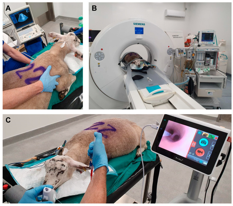
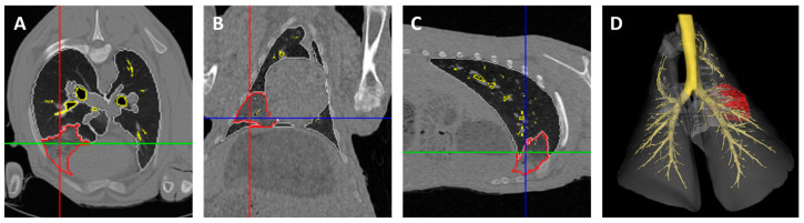
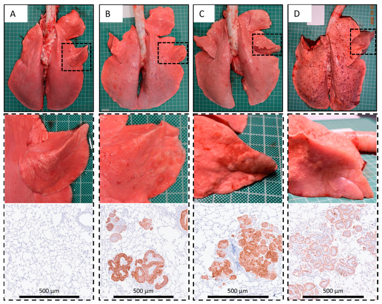
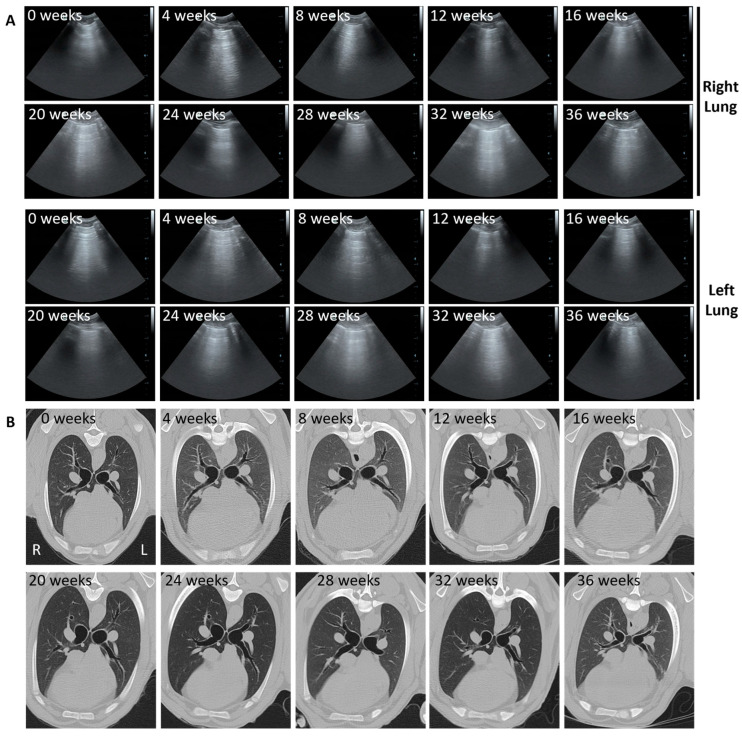
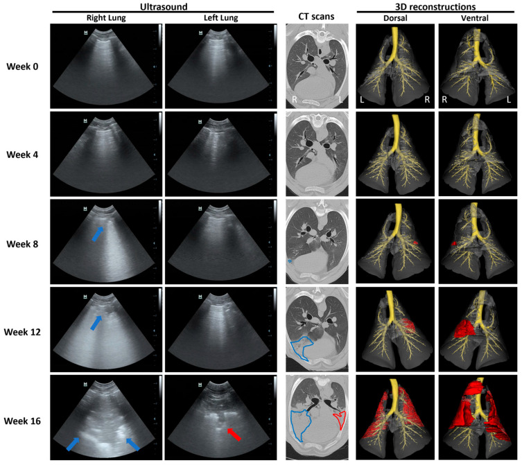
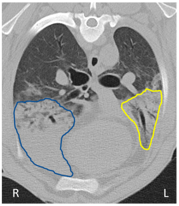
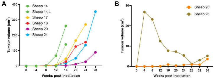
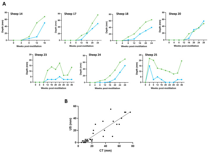
Ovine pulmonary adenocarcinoma (OPA) is an infectious, neoplastic lung disease of sheep that causes significant animal welfare and economic issues throughout the world. Understanding OPA pathogenesis is key to developing tools to control its impact. Central to this need is the availability of model systems that can monitor and track events after Jaagsiekte sheep retrovirus (JSRV) infection. Here, we report the development of an experimentally induced OPA model intended for this purpose. Using three different viral dose groups (low, intermediate and high), localised OPA tumour development was induced by bronchoscopic JSRV instillation into the segmental bronchus of the right cardiac lung lobe. Pre-clinical OPA diagnosis and tumour progression were monitored by monthly computed tomography (CT) imaging and trans-thoracic ultrasound scanning. Post mortem examination and immunohistochemistry confirmed OPA development in 89% of the JSRV-instilled animals. All three viral doses produced a range of OPA lesion types, including microscopic disease and gross tumours; however, larger lesions were more frequently identified in the low and intermediate viral groups. Overall, 31% of JSRV-infected sheep developed localised advanced lesions. Of the sheep that developed localised advanced lesions, tumour volume doubling times (calculated using thoracic CT 3D reconstructions) were 14.8 ± 2.1 days. The ability of ultrasound to track tumour development was compared against CT; the results indicated a strong significant association between paired CT and ultrasound measurements at each time point (R2 = 0.799, p < 0.0001). We believe that the range of OPA lesion types induced by this model replicates aspects of naturally occurring disease and will improve OPA research by providing novel insights into JSRV infectivity and OPA disease progression.

Keywords: animal models; computed tomography; ovine pulmonary adenocarcinoma; ultrasound.


Conflict of interest statement

The authors declare that C.C. and M.G. received a research grant from Johnson & Johnson to complete this study. J.S., N.S., and C.E.E. are employees of Johnson & Johnson. The funders had no role in the collection, analysis, or interpretation of data or in the decision to publish the results. C.E.E. made critical revisions to the manuscript. The remaining authors declare that the research was conducted in the absence of any commercial or financial relationships that could be construed as a potential conflict of interest.

Figures

Figure 1
Figure 1
Pictures documenting sheep undergoing general anaesthesia for imaging and virus instillation. (A) Trans-thoracic ultrasound, (B) CT imaging, and (C) Bronchoscopy and virus instillation.
Figure 2
Figure 2
CT image analysis for the identification of regions of interest and tumour volumes. Each CT slice was segmented to create regions of interest, including the trachea, mainstem bronchi, small airways, left and right normal lungs, and tumour areas. Blue, green and red lines indicate CT slices corresponding to the shown axial (A), coronal (B) and sagittal (C) planes. (D) Final 3D reconstruction, dorsal view. Trachea, mainstem, and segmental and subsegmental bronchi (yellow); left, right, and accessory lung lobes (grey); right lung tumour (red).
Figure 3
Figure 3
Gross post mortem images and representative IHC results. (A) Control (sheep No. 3). (B) OPA-positive: no gross lesions, JSRV positive cells detected by IHC (sheep No. 28). (C) OPA-positive: small early lesions with a width of <2 cm visible on the pleural surface, JSRV-positive cells detected by IHC (sheep No. 25). (D) OPA-positive: localised advanced lesions affecting greater than half of the affected lobe, JSRV-positive cells detected by IHC (sheep No. 20). IHC was performed using an antibody raised against the JSRV envelope SU protein. Pleural surface mottling apparent in the lungs in panel D represents post-euthanasia artefact.
Figure 4
Figure 4
Sheep weight gain over the course of the study. (A) Average sheep weight gain with respect to treatment group. Mixed-effects analysis followed by a Tukey’s multiple comparison test, comparing all of the different groups to each other at each time point (mean ± SEM, n = 3–6). (B) Average weight gain with sheep separated into groups by tumour status at the time of euthanasia. Mixed-effects analysis followed by Tukey’s multiple comparison test, comparing each of the different groups to each other at each time point (mean ± SEM, n = 2–13).
Figure 5
Figure 5
Normal thoracic ultrasound and CT appearance of ovine lungs from a control sheep over 9 months. (A) Transverse ultrasound images taken at the 4–6th intercostal spaces in the region of the cardiac lung lobes. An uninterrupted hyperechoic line consistent with the normal appearance of the visceral pleural surface can be seen at all time points. (B) Axial CT images highlighting the left and right cardiac lung lobes. Normal lung characterised by hypoattenuating lung parenchyma, through which air-filled (black) bronchi and bronchioles run, can be seen at all time points.
Figure 6
Figure 6
Serial ultrasound, CT and 3D reconstructions documenting OPA tumour development. Transverse ultrasound images were taken at the 4–6th right and left intercostal spaces in the region of the cardiac lung lobes. Blue arrows indicate progression of a right cardiac lobe OPA lesion from an irregular hyperechoic pleural line with B lines at 8 weeks to a large hypoechoic lesion at 16 weeks post-JSRV instillation. Red arrow indicates a left cardiac lobe OPA lesion identified at 16 weeks post-JSRV instillation. Axial CT images highlighting the left and right cardiac lung lobes. Blue and red lines highlight the progression of right and left OPA lesions, respectively. CT 3D reconstructions include the trachea, mainstem, segmental, and subsegmental bronchi (yellow); left, right, and accessory lung lobes (grey); left and right lung tumours (red).
Figure 7
Figure 7
CT image documenting difficulties in CT-based OPA diagnosis. Axial CT image highlighting the left and right cardiac lung lobes from sheep 17. The region outlined in blue highlights hyperattenuating lung in the instilled right cardiac lung lobe, subsequently confirmed as OPA. The region outlined in yellow highlights hyperattenuating lung in the contralateral left cardiac lung lobe that was grossly normal and demonstrated no evidence of OPA by IHC.
Figure 8
Figure 8
OPA tumour volumes. (A) Graph showing right cardiac lobe tumour volumes from all 5 sheep that were identified at post mortem examination as having advanced localised gross OPA lesions. The gross lesion that developed in the contralateral left cardiac lung lobe of sheep 14 is also shown (sheep 14 L, dashed line). (B) Graph showing tumour volumes from the 2 sheep that had gross post mortem lesions with <2 cm width visible on the pleural surface.
Figure 9
Figure 9
Changes in OPA lesion depth over time using CT and ultrasound. Measurements were taken from lesions identified within the right cardiac lung lobe. (A) Corresponding CT and ultrasound measurements were taken at each time point. CT tumour depth (green), ultrasound tumour depth (blue). (B) Pearson correlation scatterplot of paired CT and ultrasound measurements (R2 = 0.799; p < 0.0001).

Similar articles

Cited by

References

    1. DeMartini J.C., York D.F. Retrovirus-associated neoplasms of the respiratory system of sheep and goats: Ovine pulmonary carcinoma and enzootic nasal tumor. Vet. Clin. N. Am. Food Anim. Pract. 1997;13:55–70. doi: 10.1016/S0749-0720(15)30364-9. - DOI - PubMed
    1. Palmarini M., Fan H., Sharp J.M. Sheep pulmonary adenomatosis: A unique model of retrovirus associated lung cancer. Trends Microbiol. 1997;5:478–483. doi: 10.1016/S0966-842X(97)01162-1. - DOI - PubMed
    1. Griffiths D.J., Martineau H., Cousens C. Pathology and pathogenesis of ovine pulmonary adenocarcinoma. J. Comp. Pathol. 2010;142:260–283. doi: 10.1016/j.jcpa.2009.12.013. - DOI - PubMed
    1. Leroux C., Girard N., Cottin V., Greenland T., Mornex J.-F., Archer F. Jaagsiekte Sheep Retrovirus (JSRV): From virus to lung cancer in sheep. Vet. Res. 2007;38:211–228. doi: 10.1051/vetres:2006060. - DOI - PubMed
    1. Fan H. Jaagsiekte Sheep Retrovirus and Lung Cancer. Volume 275 Springer Science & Business Media; Berlin/Heidelberg, Germany: 2003.

MeSH terms

Grants and funding

LinkOut - more resources