. 2024 Aug 3;15(8):1023.
doi: 10.3390/genes15081023.

ARGONAUTE2 Localizes to Sites of Sporocysts in the Schistosome-Infected Snail, Biomphalaria glabrata

Affiliations

ARGONAUTE2 Localizes to Sites of Sporocysts in the Schistosome-Infected Snail, Biomphalaria glabrata

Phong Phan et al. Genes (Basel). .

Abstract

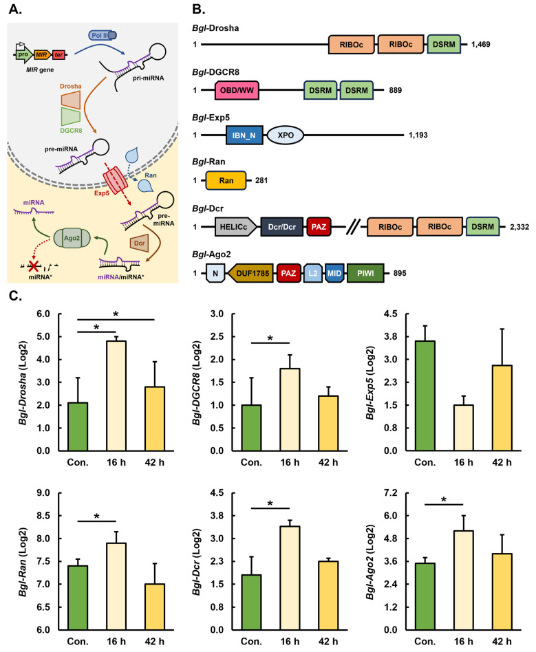
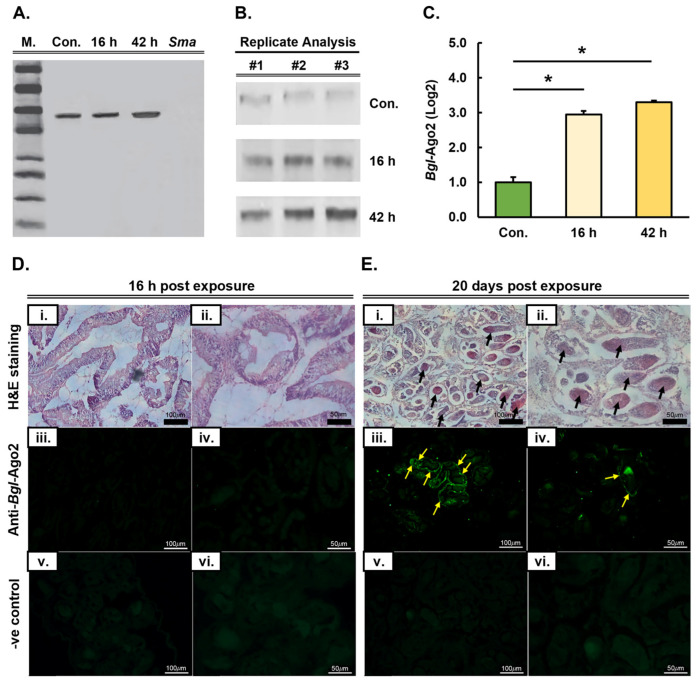
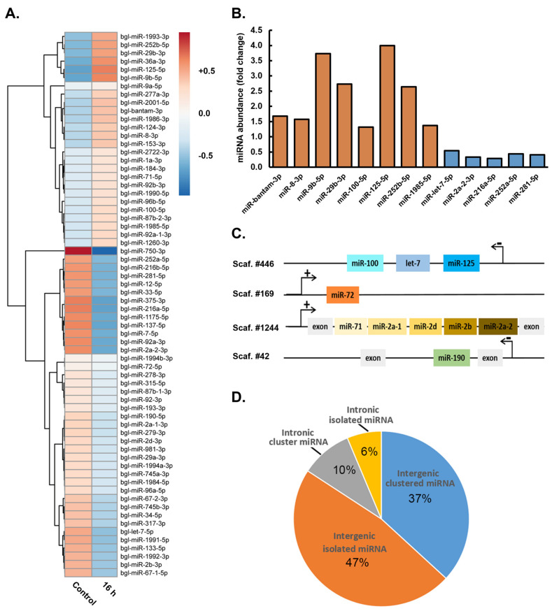
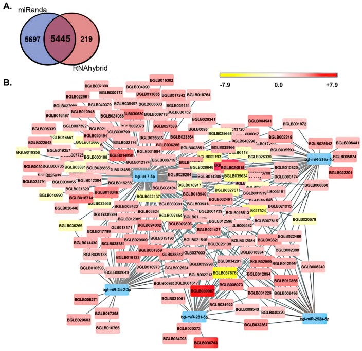
MicroRNAs (miRNAs) are a class of small regulatory RNA that are generated via core protein machinery. The miRNAs direct gene-silencing mechanisms to mediate an essential role in gene expression regulation. In mollusks, miRNAs have been demonstrated to be required to regulate gene expression in various biological processes, including normal development, immune responses, reproduction, and stress adaptation. In this study, we aimed to establishment the requirement of the miRNA pathway as part of the molecular response of exposure of Biomphalaria glabrata (snail host) to Schistosoma mansoni (trematode parasite). Initially, the core pieces of miRNA pathway protein machinery, i.e., Drosha, DGCR8, Exportin-5, Ran, and Dicer, together with the central RNA-induced silencing complex (RISC) effector protein Argonaute2 (Ago2) were elucidated from the B. glabrata genome. Following exposure of B. glabrata to S. mansoni miracidia, we identified significant expression up-regulation of all identified pieces of miRNA pathway protein machinery, except for Exportin-5, at 16 h post exposure. For Ago2, we went on to show that the Bgl-Ago2 protein was localized to regions surrounding the sporocysts in the digestive gland of infected snails 20 days post parasite exposure. In addition to documenting elevated miRNA pathway protein machinery expression at the early post-exposure time point, a total of 13 known B. glabrata miRNAs were significantly differentially expressed. Of these thirteen B. glabrata miRNAs responsive to S. mansoni miracidia exposure, five were significantly reduced in their abundance, and correspondingly, these five miRNAs were determined to putatively target six genes with significantly elevated expression and that have been previously associated with immune responses in other animal species, including humans. In conclusion, this study demonstrates the central importance of a functional miRNA pathway in snails, which potentially forms a critical component of the immune response of snails to parasite exposure. Further, the data reported in this study provide additional evidence of the complexity of the molecular response of B. glabrata to S. mansoni infection: a molecular response that could be targeted in the future to overcome parasite infection and, in turn, human schistosomiasis.

Keywords: Argonaute2 (Ago2); Biomphalaria glabrata; Schistosoma mansoni; host–parasite interaction; immune response; miRNA pathway protein machinery; miRNA-directed gene expression regulation; microRNA (miRNA) pathway.


Conflict of interest statement

The authors declare no conflicts of interest.

Figures

Figure 1
Figure 1
Identification and expression analysis of core pieces of protein machinery of the Biomphalaria glabrata miRNA pathway 16 and 42 h after its exposure to Schistosoma mansoni miracidia. (A) Schematic of the production (nucleus; grey-colored shaded region) and action (cytoplasm; pale-yellow-colored shaded region) stages of the B. glabrata miRNA pathway with the functional position within the pathway of Bgl-Drosha, Bgl-DGCR8, Bgl-Exp5, Bgl-Ran, Bgl-Dcr, and Bgl-Ago2 indicated. (B) Schematic outlining the functional domain structure of the core pieces of protein machinery of the B. glabrata miRNA pathway, including Bgl-Drosha, Bgl-DGCR8, Bgl-Exp5, Bgl-Ran, Bgl-Dcr, and Bgl-Ago2. RIBOc, ribonuclease III C terminal domain; DSRM, double-stranded RNA motif; OBD/WW, origin-binding domain/tryptophan-tryptophan domain; XPO, Exportin domain; IBN_N, Importin-β N-terminal domain; Ran, Ras-related nuclear domain; Dcr/Dcr, Dicer dimerization domain, HELICc, helicase superfamily C-terminal domain; PAZ, PIWI, Argonaute, and Zwille domain; N, Ago protein amino-terminal region domain; DUF1785, domain of unknown function 1785 domain; PIWI, P-element-induced wimpy testis domain; L2, linker region 2; MID, middle domain. (C) RNA-Seq assessment of the altered expression of Bgl-Drosha, Bgl-DGCR8, Bgl-Exp5, Bgl-Ran, Bgl-Dcr, and Bgl-Ago2, 16 and 42 h after exposure of B. glabrata animals to the S. mansoni parasite. Error bars represent the standard error of the mean, and an asterisk (*) denotes significantly altered transcript abundance (p-value ≤ 0.05) at either the 16 or 42 h time point compared to the level of expression of each core piece of protein machinery in control (unexposed) B. glabrata animals.
Figure 2
Figure 2
Expression analysis and tissue localization of the Ago2 protein in Biomphalaria glabrata animals following their short- and long-term exposure to the Schistosoma mansoni parasite. (A) Western blot hybridization analysis of Ago2 protein abundance in whole-protein extracts from unexposed (control) and exposed B. glabrata whole animals and S. mansoni miracidia. (B) Western blot hybridization analysis of Ago2 protein abundance in unexposed B. glabrata whole animals (control) and in exposed animals 16 and 42 h after their exposure to S. mansoni miracidia, an analysis that was performed in triplicate for quantification (C) to definitively demonstrate significantly altered Ago2 protein abundance in S. mansoni-exposed B. glabrata whole animals. An asterisk (*) denotes significantly altered Ago2 protein abundance (p-value ≤ 0.05) at either the 16 or 42 h time point compared to the level of Ago2 protein in control (unexposed) B. glabrata animals. (D) Light-field and fluorescent microscopic analysis of the intestinal digestive gland of sectioned B. glabrata animals 16 h post exposure to S. mansoni miracidia. Specifically, (Di,Dii) show different magnifications of bright-field microscopic analysis of H&E-stained regions of the B. glabrate intestinal digestive gland. No fluorescence was observed in the intestinal digestive gland of sectioned B. glabrata animals 16 h post exposure to S. mansoni miracidia (Diii,Div), nor were fluorescent signals observed in negative control samples (Dv,Dvi). (E) Light-field microscopic analysis and fluorescent microscopic analysis of the intestinal digestive gland of sectioned B. glabrata animals 20 days post exposure to the S. mansoni parasite. Specifically, (Ei,Eii) show different magnifications of bright-field microscopic analysis of H&E-stained regions of the intestinal digestive gland of sectioned B. glabrate animals, with the black arrows indicating sporocysts. Readily observable fluorescence was observed in the intestinal digestive gland of sectioned B. glabrata animals (yellow arrows), specifically around the sporocysts that had formed at this long-term exposure time point (Eiii,Eiv). As shown in (Dv,Dvi), no fluorescence was observed in the intestinal digestive gland sectioned of B. glabrata animals 20 days post exposure to S. mansoni miracidia in the negative control samples (Eiv,Ev).
Figure 3
Figure 3
Profiling of the microRNA landscape of Biomphalaria glabrata whole animals 16 h after their exposure to Schistosoma mansoni miracidia. (A) Heat map of the 66 miRNAs with altered abundance (up- (orange) or down-regulated (blue) abundance) 16 h post exposure of B. glabrata to S. mansoni miracidia, with the intensity of shading of each tile indicating the degree of change to miRNA abundance. (B) Elevated (n = 8) or reduced (n = 5) levels of the 13 miRNAs with significantly altered abundance 16 h post exposure of B. glabrata to S. mansoni miracidia, with orange-colored columns showing up-regulated miRNAs and blue-colored columns representing down-regulated miRNAs. (C) Schematic demonstrating that miRNA abundance was altered in B. glabrata whole animals after their exposure to S. mansoni miracidia regardless of the genomic context of their encoding gene, with altered miRNAs originating from MIR gene clusters or positioned at isolated MIR gene loci in both intragenic and intergenic genomic contexts. (D) Pie chart outlining the genomic context of MIR genes from which the 66 miRNAs with altered abundance 16 h post the exposure of B. glabrata whole animals to S. mansoni miracidia are derived.
Figure 4
Figure 4
Interaction map of the putative target genes of the five Biomphalaria glabrata miRNAs with significantly reduced abundance 16 h post exposure to Schistosoma mansoni miracidia. (A) Venn diagram showing the number of predicted target genes for the five significantly reduced B. glabrata miRNAs 16 h post exposure to S. mansoni miracidia following target gene assessment using the miRanda and RNAhybrid prediction tools, respectively. (B) miRNA/target gene interaction map for the five significantly reduced B. glabrata miRNAs (blue blocks) post exposure to S. mansoni miracidia, including putative target genes both with down-regulated (yellow blocks) and up-regulated (red blocks) levels of expression.

Similar articles

Cited by

References

    1. Huang J., Luo X., Huang M., Liu G., You W., Ke C. Identification and characteristics of muscle growth-related microRNA in the Pacific abalone, Haliotis discus hannai. BMC Genom. 2018;19:915. doi: 10.1186/s12864-018-5347-9. - DOI - PMC - PubMed
    1. Walker S.E., Spencer G.E., Necakov A., Carlone R.L. Identification and Characterization of microRNAs during Retinoic Acid-Induced Regeneration of a Molluscan Central Nervous System. Int. J. Mol. Sci. 2018;19:2741. doi: 10.3390/ijms19092741. - DOI - PMC - PubMed
    1. Xu F., Wang X., Feng Y., Huang W., Wang W., Li L., Fang X., Que H., Zhang G. Identification of conserved and novel microRNAs in the Pacific oyster Crassostrea gigas by deep sequencing. PLoS ONE. 2014;9:e104371. doi: 10.1371/journal.pone.0104371. - DOI - PMC - PubMed
    1. Zhao X., Yu H., Kong L., Liu S., Li Q. High throughput sequencing of small RNAs transcriptomes in two Crassostrea oysters identifies microRNAs involved in osmotic stress response. Sci. Rep. 2016;6:22687. doi: 10.1038/srep22687. - DOI - PMC - PubMed
    1. Jiao Y., Zheng Z., Du X., Wang Q., Huang R., Deng Y., Shi S., Zhao X. Identification and characterization of microRNAs in pearl oyster Pinctada martensii by Solexa deep sequencing. Mar. Biotechnol. 2014;16:54–62. doi: 10.1007/s10126-013-9528-x. - DOI - PubMed

LinkOut - more resources