. 2024 Aug 15;15(8):1080.
doi: 10.3390/genes15081080.

Activation of the CDK7 Gene, Coding for the Catalytic Subunit of the Cyclin-Dependent Kinase (CDK)-Activating Kinase (CAK) and General Transcription Factor II H, by the Trans-Activator Protein Tax of Human T-Cell Leukemia Virus Type-1

Affiliations

Activation of the CDK7 Gene, Coding for the Catalytic Subunit of the Cyclin-Dependent Kinase (CDK)-Activating Kinase (CAK) and General Transcription Factor II H, by the Trans-Activator Protein Tax of Human T-Cell Leukemia Virus Type-1

Mashiro Shirasawa et al. Genes (Basel). .

Abstract

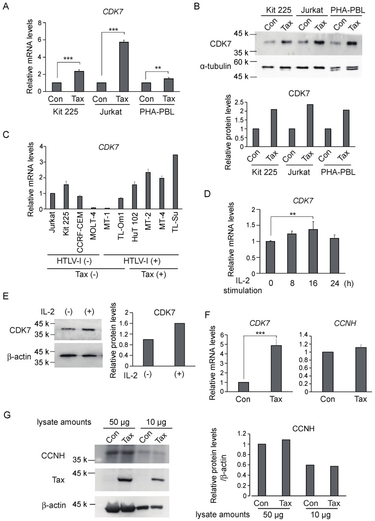
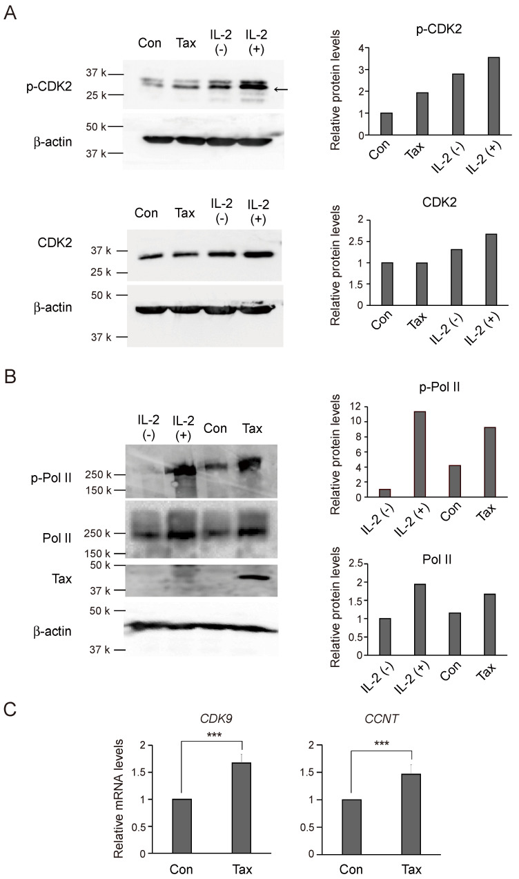
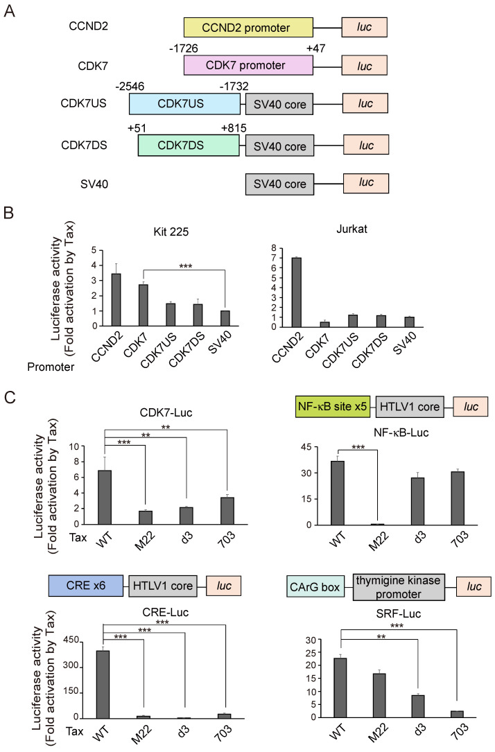
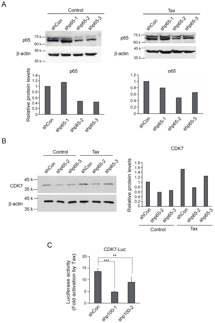
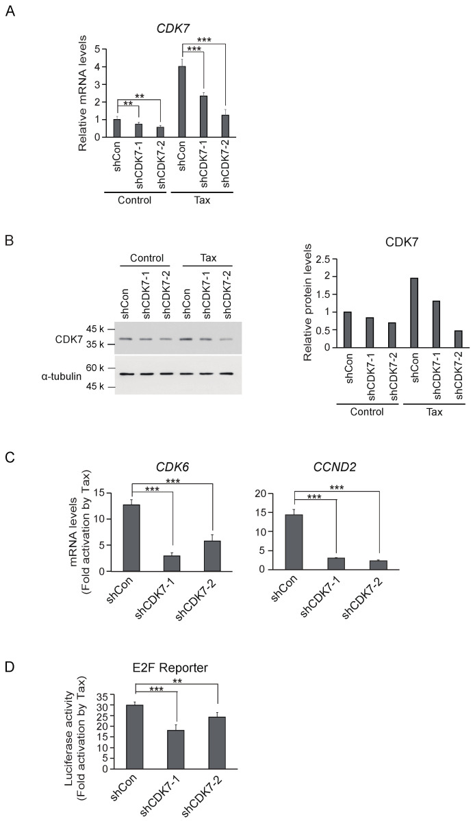
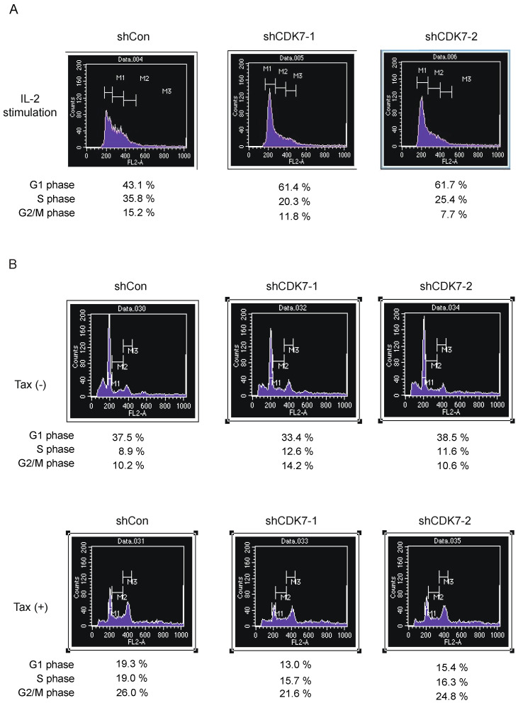
Human T-cell leukemia virus type-1 (HTLV-1) is the etiological agent of adult T-cell leukemia (ATL). The trans-activator protein Tax of HTLV-1 plays crucial roles in leukemogenesis by promoting proliferation of virus-infected cells through activation of growth-promoting genes. However, critical target genes are yet to be elucidated. We show here that Tax activates the gene coding for cyclin-dependent kinase 7 (CDK7), the essential component of both CDK-activating kinase (CAK) and general transcription factor TFIIH. CAK and TFIIH play essential roles in cell cycle progression and transcription by activating CDKs and facilitating transcriptional initiation, respectively. Tax induced CDK7 gene expression not only in human T-cell lines but also in normal peripheral blood lymphocytes (PHA-PBLs) along with increased protein expression. Tax stimulated phosphorylation of CDK2 and RNA polymerase II at sites reported to be mediated by CDK7. Tax activated the CDK7 promoter through the NF-κB pathway, which mainly mediates cell growth promotion by Tax. Knockdown of CDK7 expression reduced Tax-mediated induction of target gene expression and cell cycle progression. These results suggest that the CDK7 gene is a crucial target of Tax-mediated trans-activation to promote cell proliferation by activating CDKs and transcription.

Keywords: ATL; CDK7; HTLV-1; Tax; trans-activation.


Conflict of interest statement

The authors declare no conflicts of interest.

Figures

Figure 1
Figure 1
(A) Asynchronously growing Kit 225, Jurkat and PHA-PBLs were infected with Tax-expressing adenovirus or control virus at an MOI of 100, further cultured for 48 h in the absence of IL-2 and harvested. Expression levels of CDK7 mRNA were examined by qRT-PCR, adjusted by those of GAPDH as an internal control. Relative expression levels are presented as that of the control virus as 1. **: 0.01 ≤ p < 0.05, ***: p < 0.01. (B) Under the same condition, expression levels of CDK7 protein were examined by Western blot analysis (upper panel). α-tubulin was used as an internal control. Intensities of the bands were measured by ImageJ, and the levels of CDK7 expression were adjusted by those of α-tubulin (lower panel). (C) The expression levels of CDK7 mRNA were examined by qRT-PCR in the indicated cell lines, adjusted by those of GAPDH as an internal control. Relative expression levels are presented. (D) Kit 225 cells were starved of IL-2 for 2 days, re-stimulated by IL-2 for the indicated times and harvested. The levels of CDK7 mRNA were examined by qRT-PCR, adjusted by those of GAPDH as an internal control. Fold inductions by IL-2 are presented. **: 0.01 ≤ p < 0.05. (E) Under the same conditions, CDK7 protein expression was examined at 16 h after IL-2 stimulation by Western blot analysis (upper panel). β-actin was used as an internal control. Intensities of the bands were measured by ImageJ, and the levels of CDK7 expression were adjusted by those of β-actin (lower panel). (F) Asynchronously growing Kit 225 cells were infected with Tax-expressing adenovirus or control virus at an MOI of 100, further cultured for 48 h in the absence of IL-2 and harvested. Expression levels of CDK7 and CCNH mRNAs were examined by qRT-PCR, adjusted by those of GAPDH as an internal control. Relative expression levels are presented as that of the control virus as 1. ***: p < 0.01. (G) Under the same conditions, the level of CCNH protein was examined by Western blot analysis with two different amounts of sample (left panel). β-actin was used as an internal control. Intensities of the bands were measured by ImageJ, and the levels of CDK7 expression were adjusted by those of β-actin (right panel).
Figure 2
Figure 2
(A) Asynchronously growing Kit 225 cells were infected with Tax-expressing adenovirus or control virus at an MOI of 100, further cultured for 48 h in the absence of IL-2 and harvested. IL-2 stimulation was used as a positive control. Kit 225 cells were starved of IL-2 for 2 days, re-stimulated with IL-2 for 16 h and harvested. The levels of phosphorylated CDK2 (p-CDK2) and total CDK2 (CDK2) were examined by Western blot analysis (left panels). β-actin was used as an internal control. The intensities of p-CDK2 and CDK2 were measured by ImageJ and adjusted by those of β-actin (right panels). (B) Under the same conditions, the levels of phosphorylated CTD of RNA polymerase II (p-PolII) and total CTD of RNA polymerase II (PolII) were examined by Western blot analysis (left panels). β-actin was used as an internal control and expression of Tax was also confirmed. The intensities of p-PolII and total PolII were measured by ImageJ and adjusted by those of β-actin (right panels). (C) Under the same conditions as in Figure 1F, expression levels of CDK9 and CCNT mRNAs were examined by qRT-PCR, adjusted by those of GAPDH as an internal control. Relative expression levels are presented as that of the control virus as 1. ***: p < 0.01.
Figure 3
Figure 3
(A) Schematic presentation of reporter plasmids. (B) Kit 225 or Jurkat cells were transfected with indicated reporter plasmids along with Tax expression vector or control vector, cultured for 2 days in the absence of IL-2 and harvested. Luciferase activities were measured and normalized by those of β-galactosidase as an internal control. Fold activations by Tax are presented. ***: p < 0.01. (C) Ability of Tax mutants M22, d3 and 703 was similarly examined for activation of CDK7 promoter in Kit 225 cells. Ability of the Tax mutants to activate the NF-κB, CREB and SRF pathways was confirmed using artificial promoters containing tandem repeats of each binding site. **: 0.01 ≤ p < 0.05, ***: p < 0.01.
Figure 4
Figure 4
(A) Asynchronously growing Kit 225 cells were infected with recombinant adenovirus expressing shRNA against p65 or control virus along with Tax-expressing virus or control virus, further cultured for 3 days and harvested. Expression levels of p65 protein were examined by Western blot analysis (upper panels). The intensities of p65 bands were measured by ImageJ and adjusted by those of β-actin as an internal control (lower panels). (B) Under the same conditions, expression levels of CDK7 protein were examined by Western blot analysis except for shp65-1 (left panel). The intensities of CDK7 bands were measured by ImageJ and adjusted by those of β-actin as an internal control (right panel). (C) Asynchronously growing Kit 225 cells were transfected with CDK7 reporter along with expression vector for shRNA against p100 or control and Tax expression vector or control, further cultured for 3 days and harvested. Luciferase activities were measured and adjusted by those of β-galactosidase as an internal control. Fold activations by Tax are presented. **: 0.01 ≤ p < 0.05, ***: p < 0.01.
Figure 5
Figure 5
(A) Kit 225 cells were infected with recombinant adenovirus expressing shRNA against CDK7 along with Tax-expressing virus or control virus, cultured for 3 days and harvested. mRNA levels of CDK7 were examined by qRT-PCR, adjusted by those of GAPDH as an internal control. Relative expression levels are presented. **: 0.01 ≤ p < 0.05, ***: p < 0.01. (B) Under the same conditions, the levels of CDK7 protein were examined by Western blot analysis (left panel). α-tubulin was used as an internal control. Intensities of CDK7 bands were measured by ImageJ and adjusted by those of α-tubulin (right panel). (C) Under the same conditions, the levels of CCND2 and CDK6 mRNAs were examined by qRT-PCR. Fold inductions by Tax are shown. ***: p < 0.01. (D) Asynchronously growing Kit 225 cells were transfected with E2F reporter plasmid along with expression vector for shRNA against CDK7 or control vector and expression vector for Tax or control vector, further cultured for 3 days and harvested. Luciferase activities were measured, adjusted by those of β-galactosidase as an internal control. Fold activations are shown. **: 0.01 ≤ p < 0.05, ***: p < 0.01.
Figure 6
Figure 6
(A) Asynchronously growing Kit 225 cells were infected with recombinant adenovirus expressing shRNA against CDK7 or control virus, further cultured in the absence of IL-2 for 2 days, re-stimulated with IL-2 and harvested after 20 h. The cell cycle distribution of the cells was examined by FACS analysis of DNA content. (B) Asynchronously growing Kit 225 cells were infected with recombinant adenovirus expression shRNA against CDK7 or control virus along with Tax-expressing virus or control virus, further cultured in the absence of IL-2 for 3 days and harvested. The cell cycle distribution of the cells was determined by FACS analysis of the DNA content. The percentages of cells in G0/G1, S and G2/M are presented.

Similar articles

References

    1. Hinuma Y., Nagata K., Hanaoka M., Nakai M., Matsumoto T., Kinoshita K.I., Shirakawa S., Miyoshi I. Adult T-cell leukemia: Antigen in an ATL cell line and detection of antibodies to the antigen in human sera. Proc. Natl. Acad. Sci. USA. 1981;78:6476–6480. doi: 10.1073/pnas.78.10.6476. - DOI - PMC - PubMed
    1. Uchiyama T. Human T cell leukemia virus type I (HTLV-I) and human diseases. Annu. Rev. Immunol. 1997;15:15–37. doi: 10.1146/annurev.immunol.15.1.15. - DOI - PubMed
    1. Yoshida M. Discovery of HTLV-1, the first human retrovirus, its unique regulatory mechanisms, and insights into pathogenesis. Oncogene. 2005;24:5931–5937. doi: 10.1038/sj.onc.1208981. - DOI - PubMed
    1. Forlani G., Shallak M., Accolla R.S., Romanelli M.G. HTLV-1 Infection and Pathogenesis: New Insights from Cellular and Animal Models. Int. J. Mol. Sci. 2021;22:8001. doi: 10.3390/ijms22158001. - DOI - PMC - PubMed
    1. Osame M., Usuku K., Izumo S., Ijichi N., Amitani H., Igata A., Matsumoto M., Tara M. HTLV-I associated myelopathy, a new clinical entity. Lancet. 1986;1:1031–1032. doi: 10.1016/S0140-6736(86)91298-5. - DOI - PubMed

MeSH terms

Substances

LinkOut - more resources