. 2024 Aug 17;15(8):1087.
doi: 10.3390/genes15081087.

Genome-Wide Identification of a Maize Chitinase Gene Family and the Induction of Its Expression by Fusarium verticillioides (Sacc.) Nirenberg (1976) Infection

Affiliations

Genome-Wide Identification of a Maize Chitinase Gene Family and the Induction of Its Expression by Fusarium verticillioides (Sacc.) Nirenberg (1976) Infection

Jesús Eduardo Cazares-Álvarez et al. Genes (Basel). .

Abstract

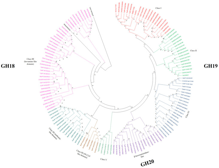
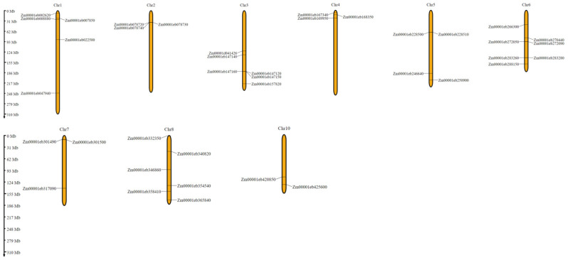
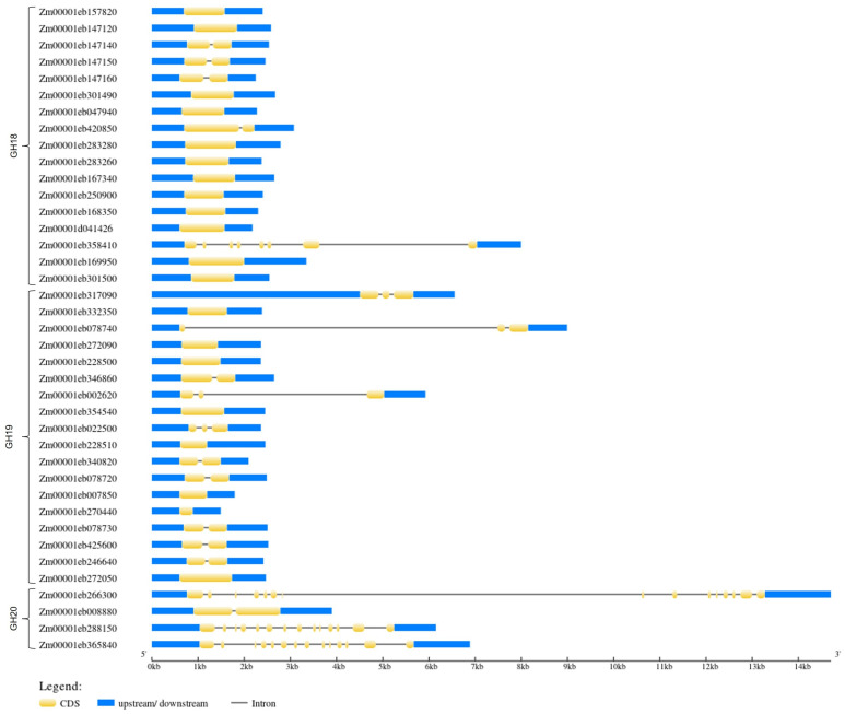
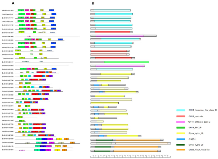
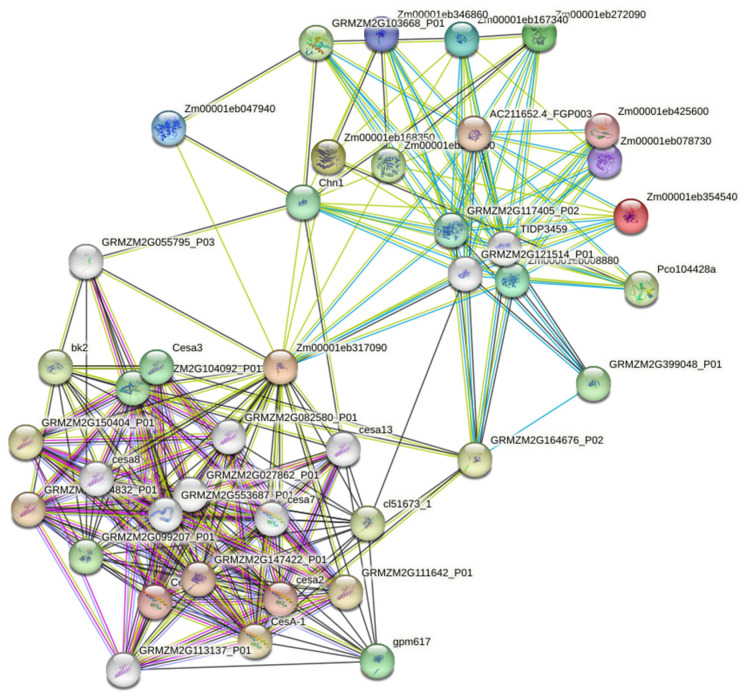
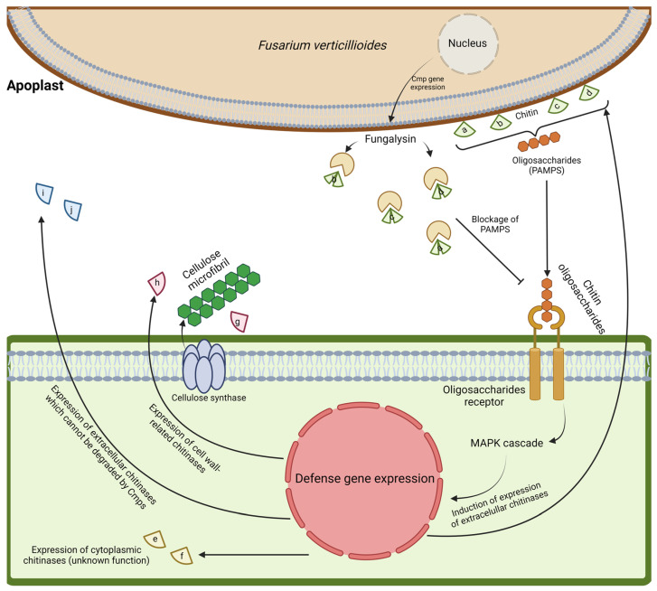
Maize chitinases are involved in chitin hydrolysis. Chitinases are distributed across various organisms including animals, plants, and fungi and are grouped into different glycosyl hydrolase families and classes, depending on protein structure. However, many chitinase functions and their interactions with other plant proteins remain unknown. The economic importance of maize (Zea mays L.) makes it relevant for studying the function of plant chitinases and their biological roles. This work aims to identify chitinase genes in the maize genome to study their gene structure, family/class classification, cis-related elements, and gene expression under biotic stress, such as Fusarium verticillioides infection. Thirty-nine chitinase genes were identified and found to be distributed in three glycosyl hydrolase (GH) families (18, 19 and 20). Likewise, the conserved domains and motifs were identified in each GH family member. The identified cis-regulatory elements are involved in plant development, hormone response, defense, and abiotic stress response. Chitinase protein-interaction network analysis predicted that they interact mainly with cell wall proteins. qRT-PCR analysis confirmed in silico data showing that ten different maize chitinase genes are induced in the presence of F. verticillioides, and that they could have several roles in pathogen infection depending on chitinase structure and cell wall localization.

Keywords: chitinase; fungi; glycosyl; hydrolase; maize; stress.


Conflict of interest statement

The authors declare no conflicts of interest.

Figures

Figure 1
Figure 1
Phylogenetic tree of Z. mays chitinase genes. GH18 includes family members of class III (hevamine-like domain; narbonin-like domain; SI-CLP-like domain); GH19 includes family members of class I, class II and class IV; and GH20 includes family members that are β-hexosaminidase-like proteins. The phylogenetic tree was built using the neighbor-joining (NJ) method and is based on the Jones–Taylor–Thornton (JTT) model, using the MUSCLE alignment function. The numbers at the tree nodes represent bootstrap percentage values. Bootstrap values are from 1000 replicates. Orthologous genes are labeled as Ta: T. aestivum; Sb: S. bicolor; Os: O. sativa.
Figure 2
Figure 2
Chitinase gene distribution in maize chromosomes. Gene names are indicated on the left and right sides of the chromosome. The Y-axis represents the distance (Mb) between genes. Their locations may be close in each chromosome, but they are not contiguous with each other.
Figure 3
Figure 3
The intron–exon structure of the chitinase-coding gene families in Z. mays. The blue bars indicate the 5′ and 3′ UTR untranslated regions, the yellow bars indicate coding sequences (CDS), and the gray lines indicate intron sequences.
Figure 4
Figure 4
Schematic structure of conserved maize chitinase motifs and domains. (A) Motifs. Colored boxes represent conserved motifs and gray lines represent non-conserved sequences; motif number sequences are shown in Table S3. (B) Domains. The conserved domains are represented by different colors.
Figure 5
Figure 5
Functional protein-association-network analysis of maize chitinases using the STRING database. Yellow lines: text mining network; black lines: co-expression; blue lines: from curated databases; violet lines: experimentally determined. Colored nodes represent the first shell of proteins interactors, and the white nodes represent the second shell of interactors.
Figure 6
Figure 6
A proposed model of maize chitinase induction in the presence of F. verticillioides. Four chitinases (a–d) possess the chitin-binding domain and Cmp site (fungalysin); these proteins may act synergistically to hydrolyze chitin in the fungal cell wall to produce chitin oligosaccharides (elicitors). These enzymes may also be the target of fungalysin, and their binding capacity could be eliminated; however, maize may produce other chitinases (i and j) that do not possess the chitin-binding domain/Cmp site to hydrolyze chitin. Due to the absence of a carbohydrate-binding domain, the chitin-binding capacity is low in these two chitinases, but their catalytic activity remains intact, and the plant could be inducing these proteins as an alternative strategy to degrade fungal chitin. On the other hand, proteins g and h, and e and f, may be induced in a coordinated mechanism to interact with cellulose synthases and to promote cell wall biosynthesis in response to Fv infection, in order to reinforce the plant cell wall. Different letters represent different chitinase proteins. Letters a–d: Zm00001eb272090, Zm00001eb346860, Zm00001eb078730, and Zm00001eb425600, respectively; letters e and f: Zm00001eb250900 and Zm00001eb168350; letters g and h: Zm00001eb317090, Zm00001eb008880; and letters i and j: Zm00001eb354540 and Zm00001eb167340.

Similar articles

Cited by

References

    1. Elieh-Ali-Komi D., Hamblin M.R. Chitin and chitosan: Production and application of versatile biomedical nanomaterials. Am. Math. Mon. 2004;111:915. - PMC - PubMed
    1. Rinaudo M. Chitin and chitosan: Properties and applications. Prog. Polym. Sci. 2006;31:603–632. doi: 10.1016/j.progpolymsci.2006.06.001. - DOI
    1. Sahraee S., Milani J.M. Chapter 8—Chitin and chitosan-based blends, composites, and nanocomposites for packaging applications. In: Gopi S., Thomas S., Pius A., editors. Handbook of Chitin and Chitosan. Elsevier; Amsterdam, The Netherlands: 2020. pp. 247–271.
    1. Aranaz I., Mengibar M., Harris R., Panos I., Miralles B., Acosta N., Galed G., Heras A. Functional characterization of chitin and chitosan. Curr. Chem. Biol. 2009;3:203–230. doi: 10.2174/187231309788166415. - DOI
    1. Lavall R.L., Assis O.B.G., Campana-Filho S.P. β-Chitin from the pens of Loligo Sp.: Extraction and characterization. Bioresour. Technol. 2007;98:2465–2472. doi: 10.1016/j.biortech.2006.09.002. - DOI - PubMed

Supplementary concepts

LinkOut - more resources