Review
. 2024 Aug 20;15(8):1095.
doi: 10.3390/genes15081095.

Genetic and Pathophysiological Basis of Cardiac and Skeletal Muscle Laminopathies

Affiliations
Review

Genetic and Pathophysiological Basis of Cardiac and Skeletal Muscle Laminopathies

Shruti Bhide et al. Genes (Basel). .

Abstract

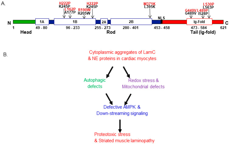
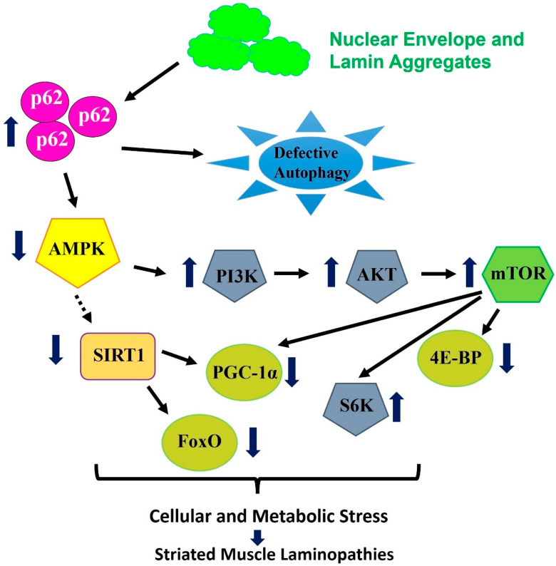
Nuclear lamins, a type V intermediate filament, are crucial components of the nuclear envelope's inner layer, maintaining nuclear integrity and mediating interactions between the nucleus and cytoplasm. Research on human iPSC-derived cells and animal models has demonstrated the importance of lamins in cardiac and skeletal muscle development and function. Mutations in lamins result in laminopathies, a group of diseases including muscular dystrophies, Hutchison-Gilford progeria syndrome, and cardiomyopathies with conduction defects. These conditions have been linked to disrupted autophagy, mTOR, Nrf2-Keap, and proteostasis signaling pathways, indicating complex interactions between the nucleus and cytoplasm. Despite progress in understanding these pathways, many questions remain about the mechanisms driving lamin-induced pathologies, leading to limited therapeutic options. This review examines the current literature on dysregulated pathways in cardiac and skeletal muscle laminopathies and explores potential therapeutic strategies for these conditions.

Keywords: Nrf2-signaling; aging; autophagy-signaling; cardiomyopathy and skeletal muscle dysfunction; laminopathies; redox-homeostasis.


Conflict of interest statement

The authors declare no conflicts of interest.

Figures

Figure 1
Figure 1
Lamin structure with mutations linked to laminopathies and crosstalk among signaling pathways. (A) Lamin structure and the location of mutated amino acids. Black: Drosophila (LamC) and red (LMNA) corresponding human numbering. The Drosophila LamC was used to depict the lamin structure with various domains. (B) The crosstalk between autophagy and redox signaling is linked with striated muscle laminopathies. The cytoplasmic aggregation of mutant LamC and other nuclear envelope protein (NEP) impairment triggers cellular and molecular stress, which leads to defective autophagy and impaired redox signaling. Impairment associated with autophagy and redox signaling leads to the inactivation of AMPK and downstream pathways, which produces proteostasis stress and leads to striated muscle laminopathy [57,62].
Figure 2
Figure 2
Model for the mechanism of AMPK/TOR/autophagy signaling by cytoplasmic lamin aggregates. Modified from Ref. [62], tissue-specific expression of lamin mutations in the heart and skeletal muscle causes the aggregation of mutant LamC and others (NEP), which causes an upregulation of p62, which ties with the aggregates and directs them for degradation [96]. Abnormal accumulation of p62 leads to autophagy inhibition in the heart and skeletal muscle, which leads to the inactivation of AMPK, which functions to maintain cellular energy homeostasis [114]. The inactivation of AMPK is associated with the hyperactivation of TOR signaling, a pathway associated with cell growth and cell survival [115,116]. In transcriptomics data obtained from muscle biopsy tissue carrying the LMNA-G449V mutation, the above-mentioned genes were upregulated, thereby leading to autophagy inhibition in the cardiac muscle [106]. The upregulation of mTOR activity also causes enhanced S6K activity and leads to an imbalance in energy homeostasis [116,117]. Moreover, AMPK is also known to regulate the expression level of genes associated with SIRT1, a class III histone deacetylase, which is linked with proteostasis and energy metabolism [110]. Furthermore, SIRT1, in turn, regulates the activity of downstream signaling, including PGC-1α, which is involved with the biogenesis of mitochondrial and FOXO control proteostasis and target 4E-BP, known to be associated with growth [112,113,118]. In addition to in vivo models, transcriptomics data obtained from human muscle tissue showed alterations in SIRT1 and other genes, thus leading to compromising proteostasis, inducing cellular stress and leading to the impairment of striated muscle pathology [57,62].

Similar articles

Cited by

References

    1. Dechat T., Adam S.A., Goldman R.D. Nuclear lamins and chromatin: When structure meets function. Adv. Enzym. Regul. 2009;49:157–166. doi: 10.1016/j.advenzreg.2008.12.003. - DOI - PMC - PubMed
    1. Goldman R.D., Gruenbaum Y., Moir R.D., Shumaker D.K., Spann T.P. Nuclear lamins: Building blocks of nuclear architecture. Genes Dev. 2002;16:533–547. doi: 10.1101/gad.960502. - DOI - PubMed
    1. Herrmann H., Bar H., Kreplak L., Strelkov S.V., Aebi U. Intermediate filaments: From cell architecture to nanomechanics. Nat. Rev. Mol. Cell Biol. 2007;8:562–573. doi: 10.1038/nrm2197. - DOI - PubMed
    1. Beck M., Hurt E. The nuclear pore complex: Understanding its function through structural insight. Nat. Rev. Mol. Cell Biol. 2017;18:73–89. doi: 10.1038/nrm.2016.147. - DOI - PubMed
    1. Adam S.A., Goldman R.D. Insights into the differences between the A- and B-type nuclear lamins. Adv. Biol. Regul. 2012;52:108–113. doi: 10.1016/j.advenzreg.2011.11.001. - DOI - PMC - PubMed

MeSH terms

LinkOut - more resources