

A Rare Case of Solitary Neurofibroma Misdiagnosed as Diabetic Foot Ulcer in the Toe Tip Region
- PMID: 39202482
- PMCID: PMC11356127
- DOI: 10.3390/medicina60081200
A Rare Case of Solitary Neurofibroma Misdiagnosed as Diabetic Foot Ulcer in the Toe Tip Region
Abstract
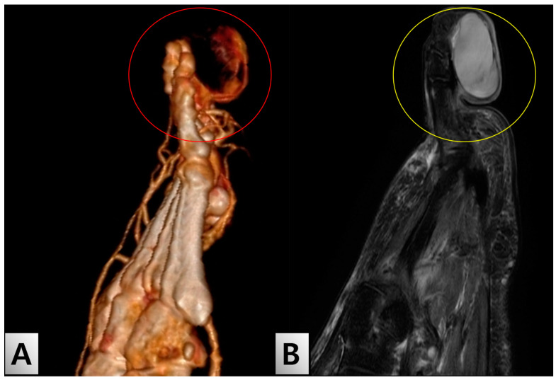
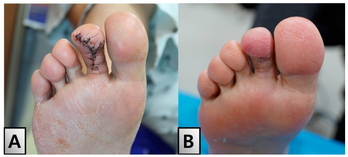
Neurofibromas, rare benign tumors of the peripheral nerve sheath, present diagnostic challenges, particularly in diabetic patients with toe ulcers. This case involves a 55-year-old female with type 2 diabetes mellitus who developed an enlarging ulcer on her right second toe. The initial evaluation suggested a diabetic ulcer; however, advanced imaging revealed a mass-like lesion. Partial excision and biopsy confirmed a neurofibroma with spindle cells within the myxoid stroma and S100 protein expression. One month later, total excision and Z-plasty reconstruction were performed under general anesthesia. The patient's postoperative recovery was uneventful, and the patient was discharged without complications. Follow-up revealed successful healing with no recurrence or functional issues. This case highlights the importance of considering neurofibromas in the differential diagnosis of diabetic toe ulcers to avoid misdiagnosis and ensure appropriate management.
Keywords: case reports; diabetes mellitus; diabetic; foot ulcer; neurofibroma; type 2.
Conflict of interest statement
The authors declare no conflicts of interest.
Figures


Similar articles
-
[Recurrence and influencing factors of diabetic foot ulcer in patients with type 2 diabetes mellitus].Zhonghua Shao Shang Za Zhi. 2020 Oct 20;36(10):947-952. doi: 10.3760/cma.j.cn501120-20190726-00315. Zhonghua Shao Shang Za Zhi. 2020. PMID: 33105947 Chinese.
-
Solitary neurofibroma of the temporal bone.J Craniofac Surg. 2010 Nov;21(6):1984-7. doi: 10.1097/SCS.0b013e3181f503be. J Craniofac Surg. 2010. PMID: 21119477
-
Rare nonsyndromic recurrent solitary gingival neurofibroma in an older adult.Clin Adv Periodontics. 2024 Jun;14(2):108-112. doi: 10.1002/cap.10260. Epub 2023 Jul 28. Clin Adv Periodontics. 2024. PMID: 37452665
-
Solitary Neurofibroma with Malignant Transformation: Case Report and Review Of Literature.Conn Med. 2015 Apr;79(4):217-9. Conn Med. 2015. PMID: 26259300 Review.
-
Hybrid Neurofibroma/Schwannoma of the Oral Cavity: A Rare Case Report and Literature Review.Int J Surg Pathol. 2023 Aug;31(5):695-701. doi: 10.1177/10668969221117978. Epub 2022 Aug 15. Int J Surg Pathol. 2023. PMID: 35971291 Review.
References
-
- Hoda S.A. Enzinger and Weiss’s Soft Tissue Tumors. Am. J. Clin. Pathol. 2020;154:424. doi: 10.1093/ajcp/aqaa078. - DOI
Publication types
MeSH terms
Substances
Grants and funding
LinkOut - more resources
Full Text Sources
Medical
