. 2024 Jul 30;12(8):1559.
doi: 10.3390/microorganisms12081559.

Lactobacillus crispatus-Mediated Gut-Reproductive Tract Axis-Alleviated Microbial Dysbiosis and Oviductal Inflammation in a Laying Hen Model

Affiliations

Lactobacillus crispatus-Mediated Gut-Reproductive Tract Axis-Alleviated Microbial Dysbiosis and Oviductal Inflammation in a Laying Hen Model

Shinuo Li et al. Microorganisms. .

Abstract

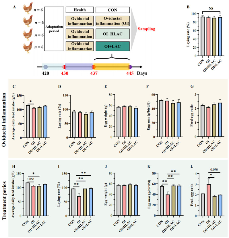
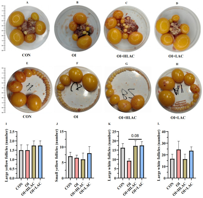
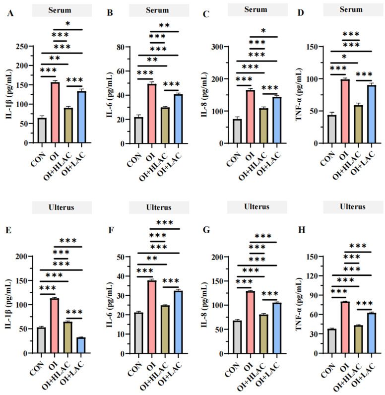
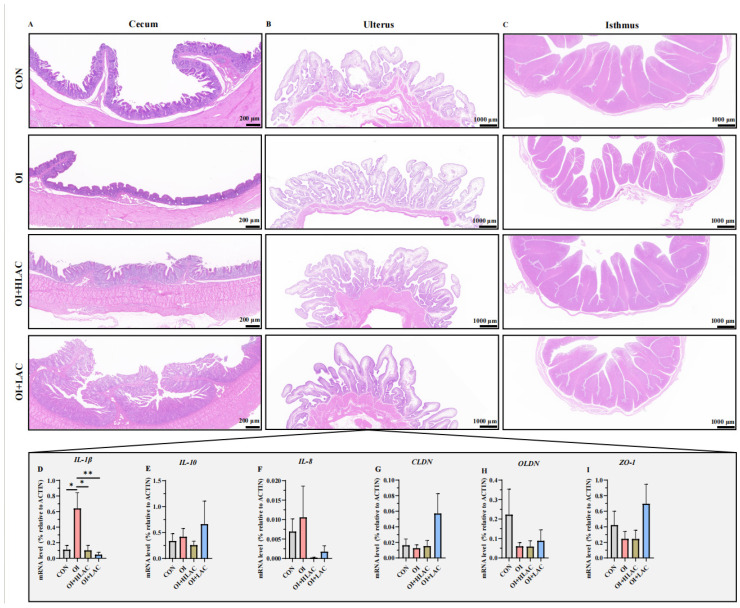
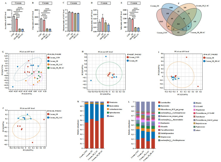
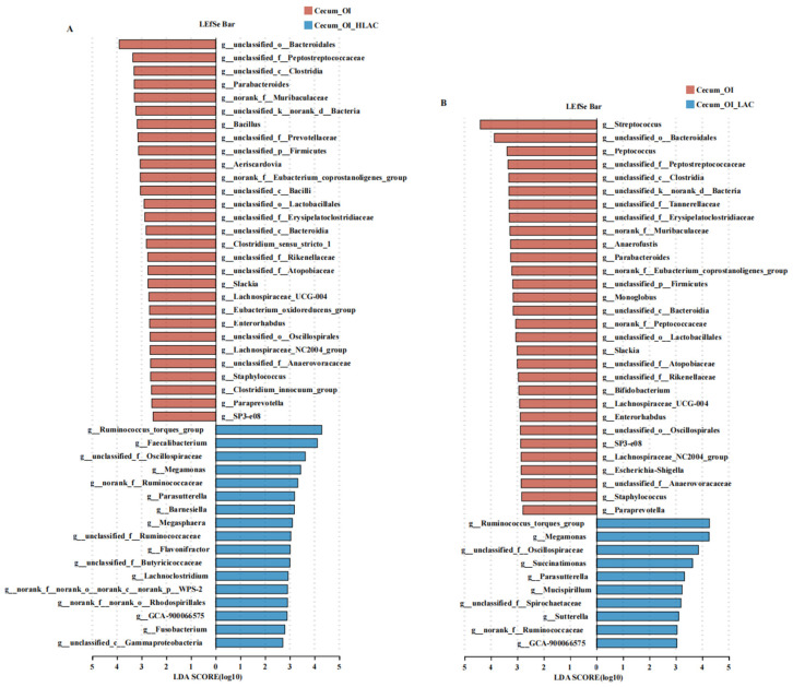
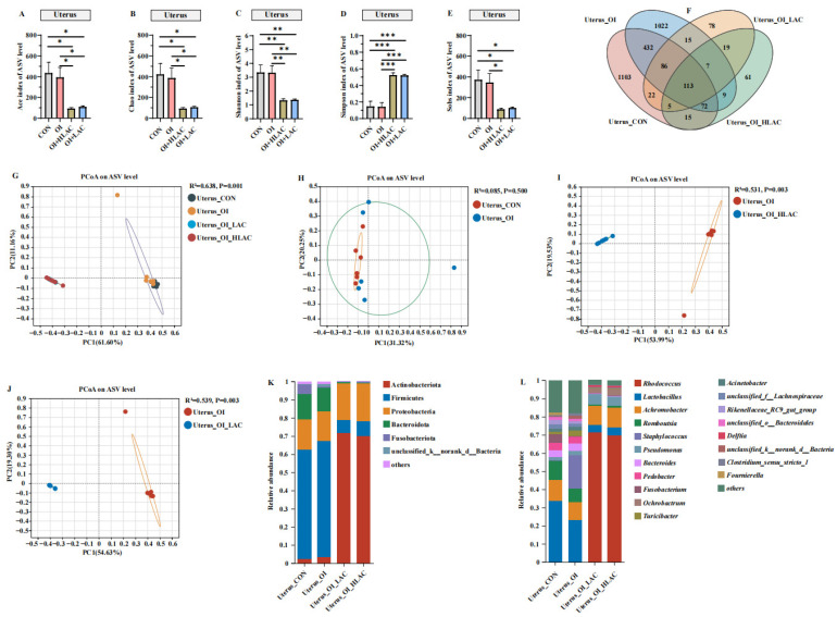
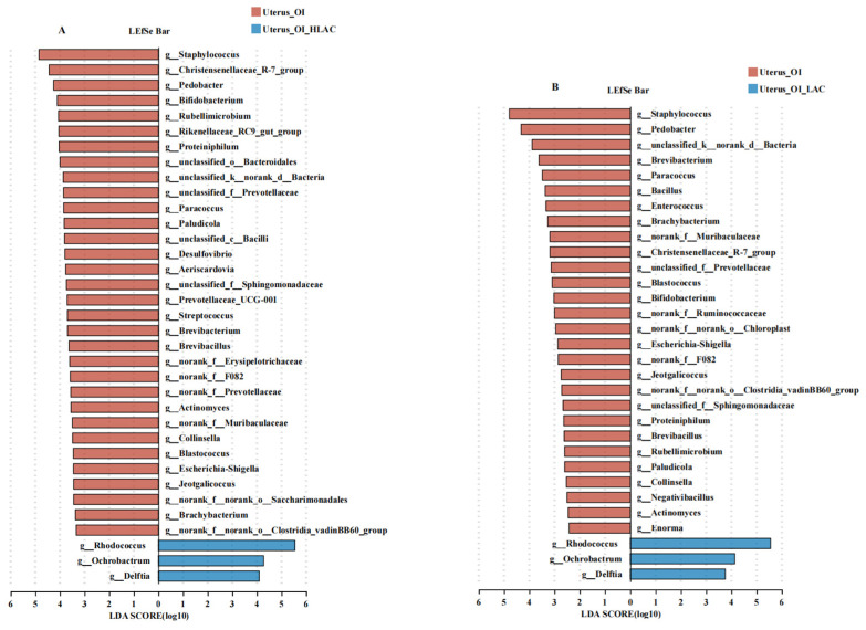
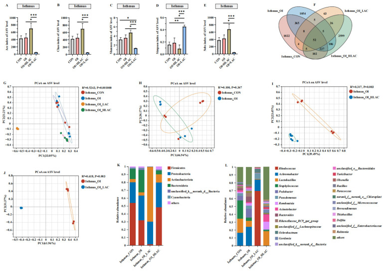
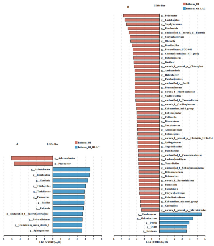
Oviductal inflammation (OI) significantly reduces the egg production and economic returns in poultry farming. While Lactobacillus crispatus (LAC) is effective against inflammation, its role in treating or preventing oviductal inflammation is understudied. In this study, we investigated the therapeutic mechanisms of LAC on oviductal inflammation, with a focus on reproductive tract health, microbiome, gene expression, and cytokine levels. This study involved 24 Jingfen No. 6 laying hens aged 60 weeks, divided into four groups: the CON, OI, OI + LAC, and OI + heat-killed Lactobacillus crispatus (HLAC) groups. And it included a 10-day adaptation, a 7-day period for the development of OI using inflammation-inducing drugs (the control received saline), followed by an 8-day treatment in which the CON and OI groups received 1 mL of MRS broth daily, and the OI + LAC and OI + HLAC groups were treated with live and heat-killed Lactobacillus crispatus (109 CFUs/mL), respectively, with six hens in each group. This study showed that Lactobacillus crispatus supplementation significantly reduced the oviductal inflammation and atrophy in the hens, with the affected hens showing markedly lower egg production rates (p < 0.001) compared to the control and treated groups (OI + HLAC and OI + LAC). The daily intake of fresh (OI + LAC, p = 0.076) or heat-killed (OI + HLAC, p < 0.01) Lactobacillus crispatus notably enhanced the feed conversion efficiency. The OI group suffered significant ovarian damage and vascular rupture, more so than the CON group, while Lactobacillus crispatus supplementation mitigated this damage. The IL-1β, IL-6, and IL-8 levels were significantly elevated in the OI group compared to those in the OI + LAC group (p < 0.05), with a significant reduction in the TNF-α levels in the latter (p < 0.001). The supplementation improved the microbial composition in the cecum, isthmus, and shell gland, enriching the cecum with beneficial bacteria, such as Ruminococcus_torques_group and Megamonas. This approach fostered ovarian health and follicle differentiation and preserved the epithelial cell barrier function in the shell gland, reducing inflammatory damage in the genital tract. This dual efficacy underscores the role of the probiotic in diminishing oviductal inflammation, regardless of its state.

Keywords: Lactobacillus crispatus; gut–reproductive tract axis; hens; laying performance; microbiome; salpingitis.


Conflict of interest statement

The authors declare no conflicts of interest.

Figures

Figure 1
Figure 1
Effects of Lactobacillus crispatus on the laying performance of laying hens. (A) Experimental design. The experiment was conducted in three phases: first, a 10-day adaptation period; second, a 7-day period for the oviductal inflammation model creation, with the control (CON) group receiving sterile saline and the oviductal inflammation (OI), OI + heat-killed Lactobacillus crispatus (HLAC), and OI + Lactobacillus crispatus (LAC) groups receiving daily inflammation-inducing drugs; and third, an 8-day treatment period during which the CON and OI groups were administered 1 mL of MRS broth daily using gavage, the OI + LAC group received fresh Lactobacillus crispatus (1 × 10⁹ CFUs mL−1), and the OI + HLAC group received heat-killed Lactobacillus crispatus (1 × 10⁹ CFUs mL−1), all administered in equivalent volumes using gavage once a day. (B) Average laying rate of laying hens during the adaptation period. (CG) Average daily feed intake (C), laying rate (D), egg weight (E), egg mass (F), and feed–egg ratio (G) of laying hens during oviductal inflammation period. (HL) Average daily feed intake (H), laying rate (I), egg weight (J), egg mass (K), and feed–egg ratio (L) during the treatment phase. Data are expressed as means ± SEMs (n  =  6). * p < 0.05, ** p < 0.01.
Figure 2
Figure 2
Effects of Lactobacillus crispatus on laying hen egg quality. (AE) Measurements of shell strength (A), egg weight (B), albumen height (C), yolk color (D), and Haugh unit (E) for laying hens during oviductal inflammation period. (FJ) Observations of shell strength (F), egg weight (G), albumen height (H), yolk color (I), and Haugh unit (J) for laying hens during treatment period. Data are expressed as means ± SEMs (n  =  10). * p < 0.05.
Figure 3
Figure 3
Effects of Lactobacillus crispatus on laying hen ovarian health during oviductal inflammation. (AD) Ovarian states of laying hens across various treatment groups. (EH) Follicle counts of varying grades in laying hens subjected to different treatments, including LYFs (large yellow follicles) (I), SYFs (small yellow follicles) (J), LWFs (large white follicles) (K), and SWFs (small white follicles) (L). Data are shown as means ± SEMs (n  =  6).
Figure 4
Figure 4
Effects of Lactobacillus crispatus on inflammatory cytokines in laying hen serum and uterus during oviductal inflammation. (AD) IL-1β (A), IL-6 (B), IL-8 (C), and TNF-α (D) levels in laying hens during oviductal inflammation period. (EH) IL-1β (E), IL-6 (F), IL-8 (G), and TNF-α (H) levels in laying hens during treatment period. IL-1β, interleukin-1β; IL-6, interleukin-6; IL-8, interleukin-8; TNF-α, tumor necrosis factor-α. Data are expressed as means ± SEMs (n  =  6). * p < 0.05, ** p < 0.01, *** p < 0.001.
Figure 5
Figure 5
Effects of Lactobacillus crispatus on histological morphology of cecum (A), isthmus (B), and shell gland (C) tissues and inflammation-related gene expression (DI) in the shell glands of laying hens during oviductal inflammation. IL-1β, interleukin-1β; IL-10, interleukin-10; IL-8, interleukin-8; CLDN, claudin; OLDN, Occludin; ZO-1, Zonula occludens-1. Data are expressed as means ± SEMs (n  =  3). * p < 0.05, ** p < 0.01.
Figure 6
Figure 6
Effects of Lactobacillus crispatus on cecum microbiota in laying hens during oviductal inflammation. (AE) Measures of five alpha diversity metrics (Ace, Chao, Shannon, Simpson, and Sobs indices) in laying hen ceca. (F) Venn diagram illustrating Amplicon Sequence Variant (ASV) overlap. (GJ) Principal coordinate analysis (PCoA) plots based on weighted UniFrac distances depicting microbiota similarity across different groups. (K) Chart detailing average distribution of major phyla within cecal microbiota. (L) Chart presenting average proportion of predominant genera in cecal microbiota. Data are expressed as means ± SEMs (n  =  6). *** p < 0.001.
Figure 7
Figure 7
Effects of Lactobacillus crispatus on differential cecal microbes in laying hens during oviductal inflammation. (A) The linear discriminant analysis (LDA) effect size (LEfSe) analysis of the cecum microbiota between Cecum_OI (red) and Cecum_OI_HLAC (blue) groups at the genus level. (B) The LEfSe analysis of the cecum microbiota between Cecum_OI (red) and Cecum_OI_LAC (blue) groups at the genus level.
Figure 8
Figure 8
Effects of Lactobacillus crispatus on uterine microbiota of laying hens during oviductal inflammation. (AE) Measures of five alpha diversity metrics (Ace, Chao, Shannon, Simpson, and Sobs indices) in laying hen ceca. (F) Venn diagram illustrating Amplicon Sequence Variant (ASV) overlap. (GJ) Principal coordinate analysis (PCoA) plots based on weighted UniFrac distances depicting microbiota similarity across different groups. (K) Chart detailing average distribution of major phyla within cecal microbiota. (L) Chart presenting average proportion of predominant genera in cecal microbiota. Data are expressed as means ± SEMs (n  =  6). * p < 0.05, ** p < 0.01, *** p < 0.001.
Figure 9
Figure 9
Effects of Lactobacillus crispatus on differential uterine microbes of laying hens during oviductal inflammation. (A) The LEfSe analysis of the uterine microbiota between Uterus_OI (red) and Uterus_OI_HLAC (blue) groups at the genus level. (B) The LEfSe analysis of the uterine microbiota between Uterus_OI (red) and Uterus_OI_LAC (blue) groups at the genus level.
Figure 10
Figure 10
Effects of Lactobacillus crispatus on isthmus microbiota of laying hens during oviductal inflammation. (AE) Measures of five alpha diversity metrics (Ace, Chao, Shannon, Simpson, and Sobs indices) in laying hen ceca. (F) Venn diagram illustrating Amplicon Sequence Variant (ASV) overlap. (GJ) Principal coordinate analysis (PCoA) plots based on weighted UniFrac distances depicting microbiota similarity across different groups. (K) Chart detailing average distribution of major phyla within cecal microbiota. (L) Chart presenting average proportions of predominant genera in cecal microbiota. Data are expressed as means ± SEMs (n  =  6). * p < 0.05, ** p < 0.01, *** p < 0.001.
Figure 11
Figure 11
Effects of Lactobacillus crispatus on differential isthmus microbes of laying hens during oviductal inflammation. (A) The LEfSe analysis of the uterine microbiota between Isthmus_OI (red) and Isthmus_OI_HLAC (blue) groups at the genus level. (B) The LEfSe analysis of the uterine microbiota between Isthmus_OI (red) and Isthmus_OI_LAC (blue) groups at the genus level.

Similar articles

Cited by

References

    1. Landman W.J., Heuvelink A., van Eck J.H. Reproduction of the Escherichia coli peritonitis syndrome in laying hens. Avian Pathol. 2013;42:157–162. doi: 10.1080/03079457.2013.775694. - DOI - PubMed
    1. Wang C., Pors S.E., Christensen J.P., Bojesen A.M., Thøfner I. Comparison and assessment of necropsy lesions in end-of-lay laying hens from different housing systems in Denmark. Poult. Sci. 2020;99:119–128. doi: 10.3382/ps/pez569. - DOI - PMC - PubMed
    1. Fang H., Quan H., Zhang Y., Li Q., Wang Y., Yuan S., Huang S., He C. Co-Infection of Escherichia coli, Enterococcus faecalis and Chlamydia psittaci Contributes to Salpingitis of Laying Layers and Breeder Ducks. Pathogens. 2021;10:755. doi: 10.3390/pathogens10060755. - DOI - PMC - PubMed
    1. Wen C., Li Q., Lan F., Li X., Li G., Yan Y., Wu G., Yang N., Sun C. Microbiota continuum along the chicken oviduct and its association with host genetics and egg formation. Poult. Sci. 2021;100:101104. doi: 10.1016/j.psj.2021.101104. - DOI - PMC - PubMed
    1. Pors S.E., Olsen R.H., Christensen J.P. Variations in virulence of avian pathogenic Escherichia coli demonstrated by the use of a new in vivo infection model. Vet. Microbiol. 2014;170:368–374. doi: 10.1016/j.vetmic.2014.02.043. - DOI - PubMed

LinkOut - more resources