

Transplacental Infections Associated with Macavirus in Aborted Bovine Fetuses
- PMID: 39203450
- PMCID: PMC11356309
- DOI: 10.3390/microorganisms12081608
Transplacental Infections Associated with Macavirus in Aborted Bovine Fetuses
Abstract
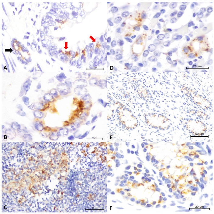
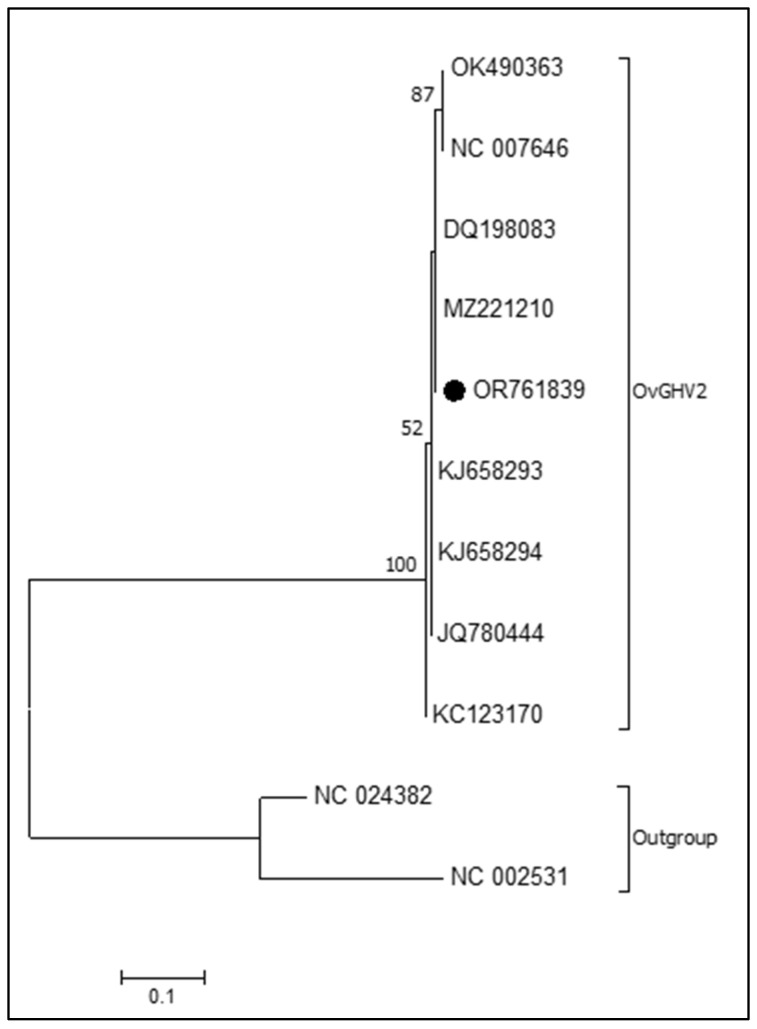
The Macavirus genus, Gammaherpesvirinae subfamily, Herpesviridae family, contains ovine gammaherpesvirus 2 (OvGHV2), the cause of sheep-associated malignant catarrhal fever (SA-MCF). Members of the Macavirus genus associated with the development of malignant catarrhal fever (MCF) in their respective hosts share the 15A antigenic epitope, are conserved within the DNA polymerase gene and are collectively referred to as the malignant catarrhal fever virus (MCFV) complex. The ability of MCFV and/or OvGHV2 to produce abortions in ruminants is currently unknown, with little documentation of infections by these agents in bovine fetuses. This report presents the findings observed due to the detection of OvGHV2 DNA and MCFV tissue antigens in aborted bovine fetuses from southern Brazil. Four aborted bovine fetuses from three farms, located in a geographical region of Paraná State with elevated immunohistochemical (IHC) prevalence of MCFV tissue antigens, with gestational ages varying between 78 to 208 days were investigated. Significant gross and histopathological alterations were not observed in any of these fetuses. An IHC assay using the 15A-monoclonal antibody (15A-MAb), which is based on the 15A antigenic epitope of Macavirus, identified MCFV tissue antigens in multiple organs from two fetuses (#1 and #4); however, positive immunoreactivity to the 15A-MAb IHC assay was not detected in Fetus #2 and #3. Molecular testing amplified OvGHV2 DNA only from the myocardium and lungs of Fetus #1 that had positive intracytoplasmic immunoreactivity to the 15A-MAb IHC assay in these tissues. Furthermore, infections by Leptospira spp. were confirmed by molecular assays in fetuses #1, #3, and #4, while PCR detected Neospora caninum in the myocardium of Fetus #2. Additionally, molecular assays to identify well-known fetopathy agents of cattle, including bovine viral diarrhea virus, bovine alphaherpesvirus 1, Histophilus somni, and Listeria monocytogenes, did not amplify the nucleic acids of these pathogens. PCR assays to identify bovine gammaherpesvirus 6 (BoGHV6), another Macavirus known to infect cattle in Brazil, were unsuccessful. These findings confirmed that the 15A-MAb IHC assay can be efficiently used to detect MCFV antigens in organs of aborted bovine fetuses. The identification of MCFV antigens with the simultaneous detection of OvGHV2 DNA confirmed that Fetus #1 was infected by OvGHV2 and added to the few descriptions of this infection in aborted fetuses of ruminants worldwide. Moreover, the IHC detection of MCFV in multiple organs of Fetus #4, without the molecular detection of OvGHV2 or BoGHV6, may suggest that this fetus was infected by a Macavirus that was not previously diagnosed in cattle herds from Brazil. These findings strongly suggest that OvGHV2 and MCFV can produce transplacental infections in cattle.
Keywords: Macavirus; diagnostic immunohistochemistry; fetal pathology; malignant catarrhal fever; vertical dissemination.
Conflict of interest statement
The authors declare no conflicts of interest.
Figures
Similar articles
-
Detection of bovine gammaherpesvirus 6 in tissues of aborted fetuses from dairy cows concomitantly infected by Histophilus somni.Microb Pathog. 2022 Aug;169:105621. doi: 10.1016/j.micpath.2022.105621. Epub 2022 Jun 7. Microb Pathog. 2022. PMID: 35688413
-
Clinical, epidemiological, and pathological findings of ovine gammaherpesvirus 2 infections in cattle from Southern Brazil.J Infect Dev Ctries. 2025 Jan 31;19(1):124-139. doi: 10.3855/jidc.19951. J Infect Dev Ctries. 2025. PMID: 39977476
-
Ovine gammaherpesvirus 2 vertical infections in sheep.Microb Pathog. 2025 May;202:107419. doi: 10.1016/j.micpath.2025.107419. Epub 2025 Feb 24. Microb Pathog. 2025. PMID: 40010654
-
Molecular Tools to Identify and Characterize Malignant Catarrhal Fever Viruses (MCFV) of Ruminants and Captive Artiodactyla.Viruses. 2022 Dec 1;14(12):2697. doi: 10.3390/v14122697. Viruses. 2022. PMID: 36560701 Free PMC article. Review.
-
Epidemiology of bovine malignant catarrhal fevers, a review.Vet Res Commun. 1981 Dec;5(2):127-42. doi: 10.1007/BF02214977. Vet Res Commun. 1981. PMID: 7048724 Review.
Cited by
-
Serological Detection of Ovine Gammaherpesvirus 2 Antibodies in Dairy Farms from Southern Brazil.Microorganisms. 2024 Dec 19;12(12):2629. doi: 10.3390/microorganisms12122629. Microorganisms. 2024. PMID: 39770831 Free PMC article.
-
Ovine gammaherpesvirus-2 associated manifestations with concomitant polioencephalomalacia, bovine alphaherpesvirus 1-related pneumonia, and caseous lymphadenitis in a sheep.Braz J Microbiol. 2025 Sep;56(3):2205-2223. doi: 10.1007/s42770-025-01682-1. Epub 2025 May 14. Braz J Microbiol. 2025. PMID: 40366576
References
-
- ICTV. International Committee on Taxonomy of Viruses Subfamily: Gammaherpesvirinae, Genus: Macavirus. [(accessed on 3 March 2024)]. Available online: https://ictv.global/report/chapter/herpesviridae/herpesviridae/macavirus.
Grants and funding
LinkOut - more resources
Full Text Sources
Miscellaneous
