. 2024 Aug 10;12(8):1634.
doi: 10.3390/microorganisms12081634.

Bacillus coagulans LMG S-24828 Impairs Candida Virulence and Protects Vaginal Epithelial Cells against Candida Infection In Vitro

Affiliations

Bacillus coagulans LMG S-24828 Impairs Candida Virulence and Protects Vaginal Epithelial Cells against Candida Infection In Vitro

Luca Spaggiari et al. Microorganisms. .

Abstract

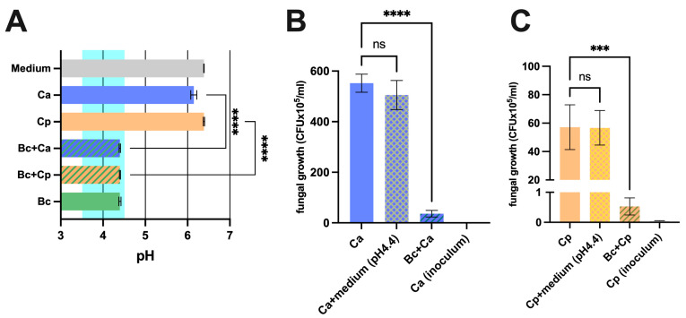
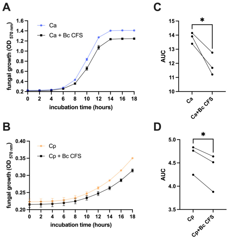
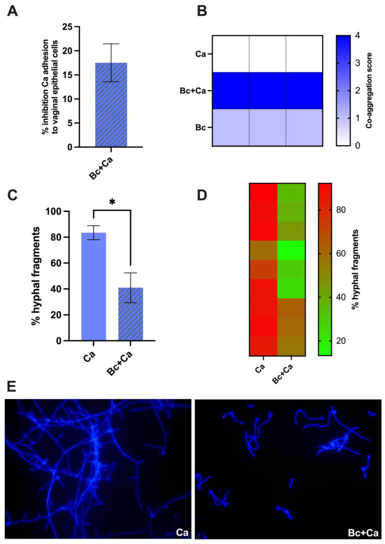
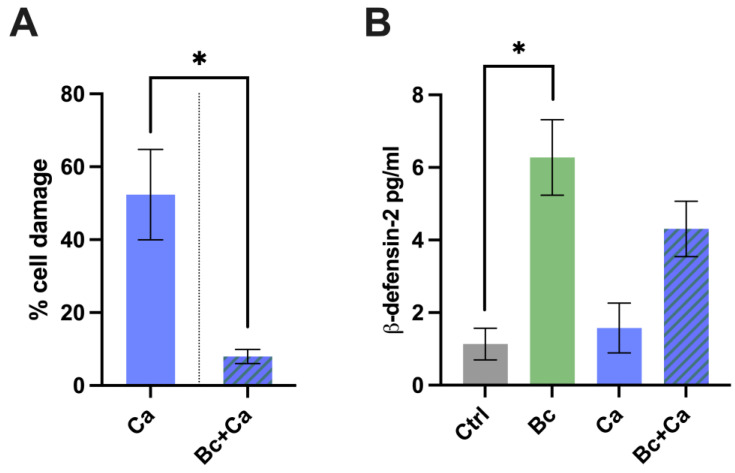
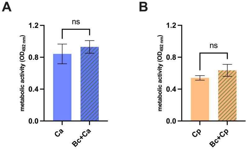
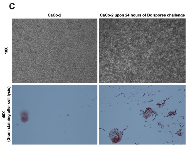
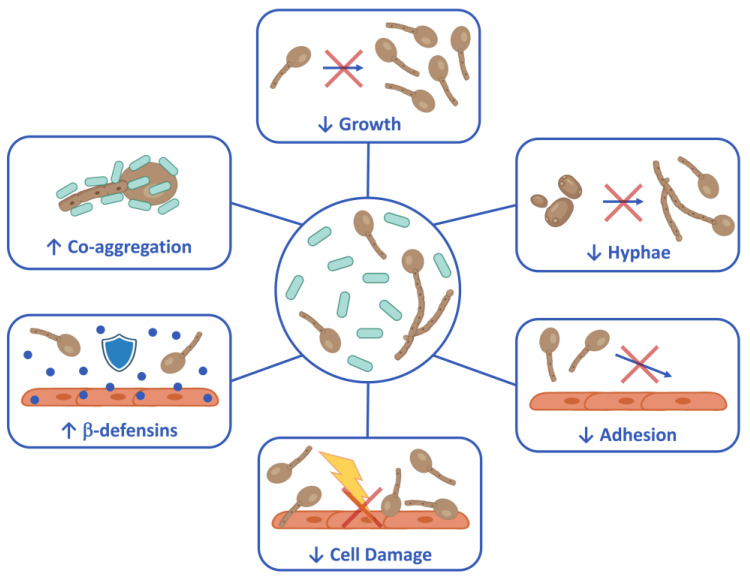
Probiotics are living microbes that provide benefits to the host. The growing data on health promotion, following probiotics administration, increased interest among researchers and pharmaceutical companies. Infections of the lower genital tract in females, caused by a wide range of pathogens, represent one of the main areas for the use of probiotics and postbiotics. Vulvovaginal candidiasis (VVC) affects 75% of women of reproductive age at least once during their lifetime, with 5-8% developing the recurrent form (RVVC). The disease is triggered by the overgrowth of Candida on the vaginal mucosa. Here, in order to establish its probiotic potential in the context of VVC, we evaluated the anti-fungal effects of the spore-producing Bacillus coagulans LMG S-24828 against C. albicans and C. parapsilosis as well as its beneficial effects in counteracting Candida vaginal infection in vitro. Our results show that both live B. coagulans and its Cell-Free Supernatant (CFS) exerted antifungal activity against both fungi. Moreover, live B. coagulans reduced hyphal formation, inhibited C. albicans adhesion to vaginal epithelial cells, showed co-aggregation capacity, and exerted a protective effect on vaginal epithelial cells infected with C. albicans. These data suggest that B. coagulans LMG S-24828 may provide benefits in the context of Candida vaginal infections.

Keywords: Bacillus coagulans; Candida; probiotics; vaginal epithelial cells.


Conflict of interest statement

Author Rosario Russo was employed by the company Giellepi S.p.A. The remaining authors declare that the research was conducted in the absence of any commercial or financial relationships that could be construed as a potential conflict of interest.

Figures

Figure 1
Figure 1
Anti-Candida effect exerted by B. coagulans. (A) Mono-cultures and co-cultures of C. albicans (Ca), C. parapsilosis (Cp), and B. coagulans (Bc) pH values after 24 h of incubation at 37 °C. The chart reports the mean values of pH ± SEM from three different experiments. The range highlighted in light green represents the mean pH levels of the healthy vaginal environment. Statistical analysis was performed by the one-way ANOVA test followed by the uncorrected Fisher’s LSD test. Ca vs. Bc + Ca **** p < 0.0001. Cp vs. Bc + Cp **** p < 0.0001. Effect of B. coagulans (Bc) on C. albicans (Ca) (B) and C. parapsilosis (Cp) (C) growth capacity and acidification contribution upon 24 h of incubation at 37 °C. The graph shows the mean C. albicans (CFU × 105/mL) ± SEM from three different experiments. Statistical analysis was performed by the one-way ANOVA test followed by the uncorrected Fisher’s LSD test. Ca vs. Bc + Ca **** p < 0.0001. Cp vs. Bc + Cp *** p < 0.001. ns = not significant.
Figure 2
Figure 2
Kinetic measurement of C. albicans (Ca) (A) or C. parapsilosis (B) growth when cultivated with B. coagulans CFS (Bc CFS) or a sterile medium at 37 °C. Culture OD at 570 nm wavelength was automatically detected every 120 min for a total of 18 h. The graphs report the mean OD values ± SEM from triplicate samples of three different experiments. The Area Under the Curve (AUC) analysis was performed on kinetic data from C. albicans (C) and C. parapsilosis (D) samples. Each line represents one single experiment. Statistical analysis was performed on AUC values through the unpaired two-tailed Student’s t-test. Ca vs. Bc CFS + Ca and Cp vs. Bc CFS + Cp * p < 0.05.
Figure 3
Figure 3
Evaluation of C. albicans (Ca) adhesion capacity to a vaginal epithelial cell monolayer in the presence of B. coagulans (Bc) (A). The histogram graph shows the average % ± SEM of fungal adhesion inhibition exerted by B. coagulans (Bc). Data are from three independent experiments. (B) Assessment of B. coagulans (Bc) capacity to co-aggregate with C. albicans (Ca) after 1 h of co-incubation. Boxes in the heatmap represent the score assigned to each sample in three independent experiments—0: no aggregation; 1: aggregates with small clusters; 2: aggregates with larger numbers of yeasts; 3: clumps visible with the naked eye containing large numbers of yeast cells; 4: maximum score for large clumps visible with the naked eye in the well center. (CE) Effect of B. coagulans (Bc) on C. albicans (Ca) hyphal formation upon 4 h of co-incubation. Hyphal fragments were optically counted by fluorescent microscopy imaging. The fungal cell wall was stained with Uvitex 2B fluorescent dye. (C) The bars chart reports the mean percentage ± SEM of hyphal fragments counted in three different fields from three independent experiments. Statistical analysis was performed by the unpaired, two-tailed Student t-test. Ca vs. Bc + Ca * p < 0.05. (D) The heatmap shows the % of hyphal fragments counted in each field; the squares’ color indicates the abundance of hyphae in the field (red: high % hyphal fragments; green: low % hyphal fragments). (E) Representative images from fluorescence microscopy analysis are shown from C. albicans (Ca) or C. albicans plus B. coagulans (Bc + Ca) taken at 40× magnification.
Figure 4
Figure 4
(A) Percentage of vaginal cell damage pre-colonized or not by B. coagulans (Bc) for 6 h and infected for further 18 h with C. albicans (Ca). The chart reports the average percentage of cell damage ± SEM of triplicate samples from three different experiments. Statistical analysis was performed by the unpaired, two-tailed Student t-test. Ca vs. Bc + Ca * p < 0.05. (B) Production of β-defensin-2 by vaginal epithelial cells pre-colonized or not by B. coagulans (Bc) for 6 h and infected for further 18 h with C. albicans (Ca). Uninfected cells (Ctrl) and cells colonized by the bacterium without C. albicans were also included in the experiments. The graph reports the mean ± SEM from three independent experiments. Statistical analysis was performed by the one-way ANOVA test followed by the uncorrected Fisher’s LSD test. Untreated cells vs. Bc pre-colonized cells * p < 0.05.
Figure 5
Figure 5
Evaluation of antifungal effect permanency upon B. coagulans removal. C. albicans (Ca) (A) and C. parapsilosis (Cp) (B) metabolic activity quantification after being incubated with B. coagulans or sterile medium for 24 h and subsequent fungal isolation and cultivation for 24 h in the lack of bacteria. The graphs show the mean OD at 492 nm wavelength ± SEM from triplicate sample of three different experiments. Statistical analysis was performed by the one-way ANOVA test followed by the uncorrected Fisher’s LSD test. ns = not significant. (C) Capacity of B. coagulans spores to germinate on intestinal epithelial cells CaCo-2. Bacterial spores were seeded on an intestinal epithelial cell monolayer of CaCo-2 and incubated at 37 °C + 5% CO2 for 24 h. After incubation, monolayers were photographed (upper images) and subsequently lysed. A Gram staining was then performed to visualize the presence of germinated B. coagulans (Bc) (lower images).
Figure 5
Figure 5
Evaluation of antifungal effect permanency upon B. coagulans removal. C. albicans (Ca) (A) and C. parapsilosis (Cp) (B) metabolic activity quantification after being incubated with B. coagulans or sterile medium for 24 h and subsequent fungal isolation and cultivation for 24 h in the lack of bacteria. The graphs show the mean OD at 492 nm wavelength ± SEM from triplicate sample of three different experiments. Statistical analysis was performed by the one-way ANOVA test followed by the uncorrected Fisher’s LSD test. ns = not significant. (C) Capacity of B. coagulans spores to germinate on intestinal epithelial cells CaCo-2. Bacterial spores were seeded on an intestinal epithelial cell monolayer of CaCo-2 and incubated at 37 °C + 5% CO2 for 24 h. After incubation, monolayers were photographed (upper images) and subsequently lysed. A Gram staining was then performed to visualize the presence of germinated B. coagulans (Bc) (lower images).
Figure 6
Figure 6
Schematic representation of biological activities of B. coagulans LMG S-24828 against Candida. Created with BioRender.com.

Similar articles

References

    1. Zhu Y.-X., Li T., Fan S.-R., Liu X.-P., Liang Y.-H., Liu P. Health-Related Quality of Life as Measured with the Short-Form 36 (SF-36) Questionnaire in Patients with Recurrent Vulvovaginal Candidiasis. Health Qual. Life Outcomes. 2016;14:65. doi: 10.1186/s12955-016-0470-2. - DOI - PMC - PubMed
    1. Sobel J.D. Recurrent Vulvovaginal Candidiasis. Am. J. Obstet. Gynecol. 2016;214:15–21. doi: 10.1016/j.ajog.2015.06.067. - DOI - PubMed
    1. Ardizzoni A., Wheeler R.T., Pericolini E. It Takes Two to Tango: How a Dysregulation of the Innate Immunity, Coupled with Candida Virulence, Triggers VVC Onset. Front. Microbiol. 2021;12:692491. doi: 10.3389/fmicb.2021.692491. - DOI - PMC - PubMed
    1. Ceccarani C., Foschi C., Parolin C., D’Antuono A., Gaspari V., Consolandi C., Laghi L., Camboni T., Vitali B., Severgnini M., et al. Diversity of Vaginal Microbiome and Metabolome during Genital Infections. Sci. Rep. 2019;9:14095. doi: 10.1038/s41598-019-50410-x. - DOI - PMC - PubMed
    1. O’Hanlon D.E., Moench T.R., Cone R.A. Vaginal pH and Microbicidal Lactic Acid When Lactobacilli Dominate the Microbiota. PLoS ONE. 2013;8:e80074. doi: 10.1371/journal.pone.0080074. - DOI - PMC - PubMed

LinkOut - more resources