. 2024 Aug 10;12(8):1640.
doi: 10.3390/microorganisms12081640.

Alteration of Trophoblast Syncytialization by Plasmodium falciparum-Infected Erythrocytes

Affiliations

Alteration of Trophoblast Syncytialization by Plasmodium falciparum-Infected Erythrocytes

Carolina López-Guzmán et al. Microorganisms. .

Abstract

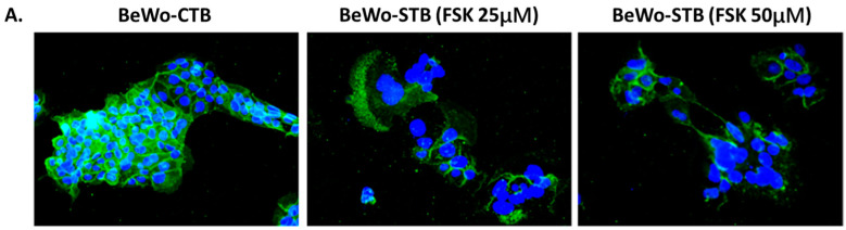
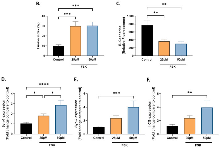
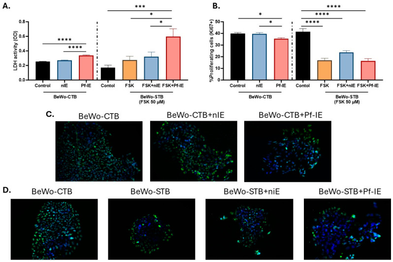
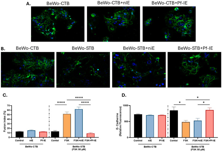
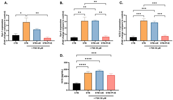
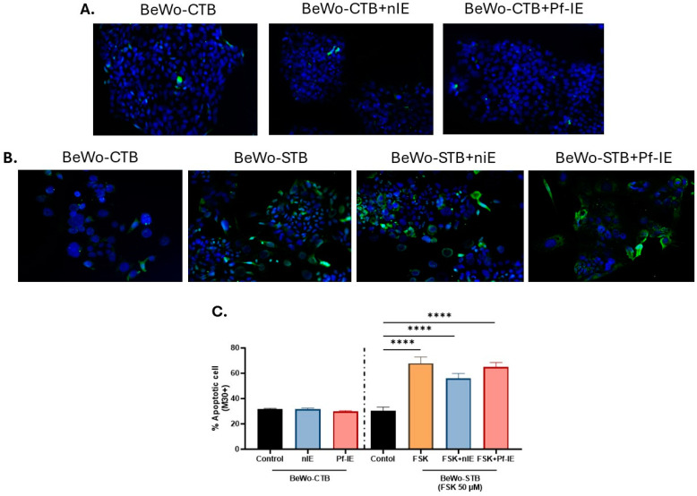
Malaria during pregnancy has been associated with significant risks to both the mother and the fetus, leading to complications such as anemia, low birth weight, and increased infant mortality. The trophoblast cells, a key component of the placenta, are crucial for nutrient and oxygen exchange between mother and fetus. The differentiation of cytotrophoblasts (CTBs) into syncytiotrophoblasts (STBs) is critical for proper pregnancy development. These cells form the bi-stratified epithelium surrounding the placental villi. While previous studies have described an inflammatory activation of STB cells exposed to Plasmodium falciparum-infected erythrocytes (P. falciparum-IE) or components such as hemozoin (HZ), little is known about the direct effect this parasite may have on the epithelial turnover and function of trophoblast cells. This study aims to contribute to understanding mechanisms leading to placental damage during placental malaria using a BeWo cell line as a differentiation model. It was found that P. falciparum-IE interferes with the fusion of BeWo cells, affecting the differentiation process of trophoblast. A reduction in syncytialization could be associated with the adverse effects of infection in fetal health, altering the remodeling of the trophoblast epithelial barrier and reducing their capacity to exchange substances. However, further studies are necessary to assess alterations in the functionality of this epithelium.

Keywords: Plasmodium falciparum; cytotrophoblast; malaria; syncytiotrophoblast; trophoblast.


Conflict of interest statement

The authors declare no conflicts of interest.

Figures

Figure 1
Figure 1
BeWo cell fusion in the presence of FSK. (A) Immunofluorescence staining for E-cadherin (green) and nuclei (blue) of cultured BeWo cells under treatment with FSK (25 μM and 50 μM, 400×). (B) Mean fluorescence intensity of E-cadherin in control cells and FSK treatments. (C) Syncytialization index in control cells and FSK treatments. (D) Expression of SYN-1 showed a 2.65-fold increase in cells treated with 50 μM FSK compared to the control. (E) Expression of SYN-2 showed a 4.04-fold increase in cells treated with 50 μM FSK compared to the control. (F) Expression of βhCG gene indicated a 3.32-fold increase in cells treated with 50 μM FSK compared to the control (n = 3). Data are presented as mean ± SEM. ANOVA, p-value: <0.00001 (****), <0.0001 (***), <0.001 (**), and <0.005 (*).
Figure 1
Figure 1
BeWo cell fusion in the presence of FSK. (A) Immunofluorescence staining for E-cadherin (green) and nuclei (blue) of cultured BeWo cells under treatment with FSK (25 μM and 50 μM, 400×). (B) Mean fluorescence intensity of E-cadherin in control cells and FSK treatments. (C) Syncytialization index in control cells and FSK treatments. (D) Expression of SYN-1 showed a 2.65-fold increase in cells treated with 50 μM FSK compared to the control. (E) Expression of SYN-2 showed a 4.04-fold increase in cells treated with 50 μM FSK compared to the control. (F) Expression of βhCG gene indicated a 3.32-fold increase in cells treated with 50 μM FSK compared to the control (n = 3). Data are presented as mean ± SEM. ANOVA, p-value: <0.00001 (****), <0.0001 (***), <0.001 (**), and <0.005 (*).
Figure 2
Figure 2
Cell viability and proliferation in BeWo cells exposed to P. falciparum-IE. (A) LDH activity was measured in the culture supernatant of BeWo-CTB and BeWo-STB cells exposed to infected erythrocytes as a marker of cytotoxicity. (B) Percentage of proliferative cells (Ki67-positive cells) in BeWo-CTB and BeWo-STB cultures exposed to P. falciparum-IE. (C) Representative images of Ki-67 (green) and nuclei (blue) immunostaining in BeWo-CTB (100X). (D) Representative images of Ki-67 (green) and nuclei (blue) immunostaining in BeWo-STB (100X). Results are presented as mean ± SEM (n = 3). Test: ANOVA, p-value: <0.00001 (****), <0.0001 (***) and <0.005 (*).
Figure 3
Figure 3
BeWo cell fusion is altered by exposure to P. falciparum-IE. Immunofluorescence staining for E-cadherin (green) and nuclei (blue) of BeWo-CTB cells (200X) (A) and BeWo-STB (200X) (B) cells under parasite treatment. (C) Fusion index under different stimulatory conditions. (D) Mean fluorescence intensity of E-cadherin different stimulatory conditions. Data are presented as mean ± SEM (n = 3). Test: ANOVA, p-value: <0.00001 (****), <0.005 (*).
Figure 4
Figure 4
Expression levels of the SYN-1, SYN-2, and βhCG genes in BeWo-STB cells under parasite treatment, evaluated by qPCR. (A) The SYN-1 gene expression. (B) The SYN-2 gene expression. (C) The βhCG gene expression. (D) hCG levels in culture supernatants BeWo cells exposed to FSK and infected erythrocytes (n = 3). Data are presented as mean ± SEM. Test: ANOVA, p-value: <0.0001 (****), <0.001 (***), <0.01 (**), <0.05 (*).
Figure 5
Figure 5
Quantification of BeWo apoptotic cells (M30-positive) exposed to P. falciparum-IE. (A) Panel of photographs of M30 staining (green) and nuclei (blue) in BeWo-CTB under different study conditions (200X). (B) Panel of photographs of M30 staining (green) and nuclei (blue) in BeWo-STB under different study conditions (200X). (C) Percentage of apoptotic cells in control cells and STB exposed or not to P. falciparum-IE. Data are expressed as mean ± SEM (n = 3). Test: ANOVA, p-value: <0.00001 (****).

Similar articles

References

    1. Rogerson S.J., Hviid L., Duffy P.E., Leke R.F., Taylor D.W. Malaria in pregnancy: Pathogenesis and immunity. Lancet Infect. Dis. 2007;7:105–117. doi: 10.1016/S1473-3099(07)70022-1. - DOI - PubMed
    1. Fried M., Duffy P.E. Malaria during Pregnancy. Cold Spring Harb. Perspect. Med. 2017;7:a025551. doi: 10.1101/cshperspect.a025551. - DOI - PMC - PubMed
    1. Moormann A.M., Sullivan A.D., Rochford R.A., Chensue S.W., Bock P.J., Nyirenda T., Meshnick S.R. Malaria and pregnancy: Placental cytokine expression and its relationship to intrauterine growth retardation. J. Infect. Dis. 1999;180:1987–1993. doi: 10.1086/315135. - DOI - PubMed
    1. Silver K.L., Zhong K., Leke R.G.F., Taylor D.W., Kain K.C. Dysregulation of angiopoietins is associated with placental malaria and low birth weight. PLoS ONE. 2010;5:e9481. doi: 10.1371/journal.pone.0009481. - DOI - PMC - PubMed
    1. Fried M., Kurtis J.D., Swihart B., Pond-Tor S., Barry A., Sidibe Y., Gaoussou S., Traore M., Keita S., Mahamar A., et al. Systemic inflammatory response to malaria during pregnancy is associated with pregnancy loss and preterm delivery. Clin. Infect. Dis. 2017;65:1729–1735. doi: 10.1093/cid/cix623. - DOI - PMC - PubMed

LinkOut - more resources