Review
. 2024 Aug 20;12(8):1713.
doi: 10.3390/microorganisms12081713.

Neurological Impact of Respiratory Viruses: Insights into Glial Cell Responses in the Central Nervous System

Affiliations
Review

Neurological Impact of Respiratory Viruses: Insights into Glial Cell Responses in the Central Nervous System

Valentina P Mora et al. Microorganisms. .

Abstract

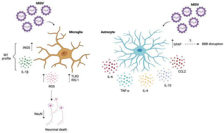
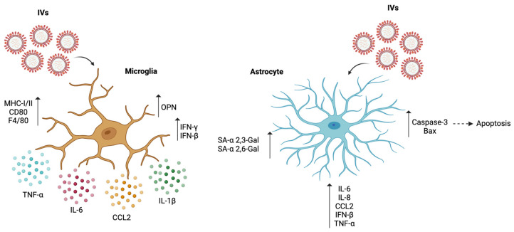
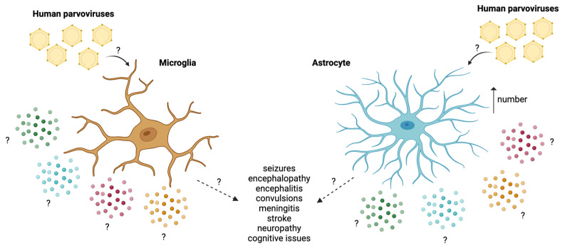
Respiratory viral infections pose a significant public health threat, particularly in children and older adults, with high mortality rates. Some of these pathogens are the human respiratory syncytial virus (hRSV), severe acute respiratory coronavirus-2 (SARS-CoV-2), influenza viruses (IV), human parvovirus B19 (B19V), and human bocavirus 1 (HBoV1). These viruses cause various respiratory symptoms, including cough, fever, bronchiolitis, and pneumonia. Notably, these viruses can also impact the central nervous system (CNS), leading to acute manifestations such as seizures, encephalopathies, encephalitis, neurological sequelae, and long-term complications. The precise mechanisms by which these viruses affect the CNS are not fully understood. Glial cells, specifically microglia and astrocytes within the CNS, play pivotal roles in maintaining brain homeostasis and regulating immune responses. Exploring how these cells interact with viral pathogens, such as hRSV, SARS-CoV-2, IVs, B19V, and HBoV1, offers crucial insights into the significant impact of respiratory viruses on the CNS. This review article examines hRSV, SARS-CoV-2, IV, B19V, and HBoV1 interactions with microglia and astrocytes, shedding light on potential neurological consequences.

Keywords: astrocytes; central nervous system; glial cells; microglia; respiratory viral infections.


Conflict of interest statement

The authors declare no conflicts of interest.

Figures

Figure 1
Figure 1
Effects of hRSV on microglia and astrocytes. The cell responses of microglia after hRSV infection indicate a polarization towards the M1 pro-inflammatory profile, alongside increases in TLR3 and RIG-1 expression. There is also an increase in ROS production, leading to neuronal death. The cell responses of astrocytes after hRSV infection led to an activation of astrocytes and BBB disruption by an unknown mechanism, accompanied by increases in the gene expression of IL-4, IL-10, and CCL2 and IL-6 and TNF-α production. Created with Biorender; Agreement #EL26KC08YQ.
Figure 2
Figure 2
Effect of SARS-CoV-2 on microglia and astrocytes. The cell responses of microglia after SARS-CoV-2 infection indicate a polarization towards an M1 pro-inflammatory profile and an increase in apoptosis, ER stress response, NLRP3 inflammasome components, IFN signaling, possibly leading to synapsis elimination and neurodegeneration. The infection also causes TLR2 activation, which is associated with depression and dysosmia. The cell responses of astrocytes after SARS-CoV-2 infection led to an activation of astrocytes, an increase in MMP9, morphological changes, and evidence of Evans blue dye leakage, suggesting a BBB disruption. There is also an increase in the IFN-I pathway, CXCL10, CXCL6, CXCL2, CXCL1, and IL-17, alongside the secretion of pro-inflammatory components TNF-α, IL-6, and CCL2. There is an increase in apoptosis and metabolic abnormalities, alongside a decrease in CAMK2D, ERBB2, C1QL, and SYPL1, which suggests neurological complications, synapse dysfunction, and neuronal death. (Created with Biorender; Agreement number #TC26KC0CKS).
Figure 3
Figure 3
Effect of IVs on microglia and astrocytes. The cell responses of microglia after IVs infection indicate an increase in MHC-I, MHC-II, and F4/80, accompanied by a pro-inflammatory profile and the secretion of TNF-α, IL-6, CCL2, and IL-1β. There is also an increase in IFN-γ, IFN-β and OPN levels. The cell responses of astrocytes after IVs infection are characterized by an increase in the expression of caspase-3 and Bax, leading to an increase in apoptosis. It also shows a pro-inflammatory profile with increased IL-6, IL-8, CCL2, IFN-β and TNF-α. There is also an increase in the expression of IVs receptors SA-α 2,3-Gal and SA-α 2,6-Gal. (Created with Biorender; Agreement number #HF26KDBRMQ).
Figure 4
Figure 4
Effect of B19V and HBoV1 on microglia and astrocytes. It is unknown how parvoviruses interact with microglia and astrocytes, and different cytokines may be produced (interrogation points). It has been described that B19V-positive astrocytes increase after infection. Neurological sequelae associated with human parvoviruses include seizures, encephalopathy, encephalitis, convulsions, meningitis, stroke, neuropathy, and cognitive issues; however, it is unknown how B19V and HBoV1 cause these effects. (Created with Biorender; Agreement number #UN26KDEMUO).

References

    1. Jessen K.R. Glial cells. Int. J. Biochem. Cell Biol. 2004;36:1861–1867. doi: 10.1016/j.biocel.2004.02.023. - DOI - PubMed
    1. Allen N.J., Lyons D.A. Glia as architects of central nervous system formation and function. Science. 2018;362:181–185. doi: 10.1126/science.aat0473. - DOI - PMC - PubMed
    1. Xu S., Lu J., Shao A., Zhang J.H., Zhang J. Glial Cells: Role of the Immune Response in Ischemic Stroke. Front. Immunol. 2020;11:294. doi: 10.3389/fimmu.2020.00294. - DOI - PMC - PubMed
    1. Bohmwald K., Gálvez N.M.S., Ríos M., Kalergis A.M. Neurologic alterations due to respiratory virus infections. Front. Cell. Neurosci. 2018;12:386. doi: 10.3389/fncel.2018.00386. - DOI - PMC - PubMed
    1. Heegaard E.D., Brown K.E. Human parvovirus B19. Clin. Microbiol. Rev. 2002;15:485–505. doi: 10.1128/CMR.15.3.485-505.2002. - DOI - PMC - PubMed

LinkOut - more resources