. 2024 Aug 9;12(8):903.
doi: 10.3390/vaccines12080903.

Long-Term Protective Immunity against Ehrlichia chaffeensis Infection Induced by a Genetically Modified Live Vaccine

Affiliations

Long-Term Protective Immunity against Ehrlichia chaffeensis Infection Induced by a Genetically Modified Live Vaccine

Swetha Madesh et al. Vaccines (Basel). .

Abstract

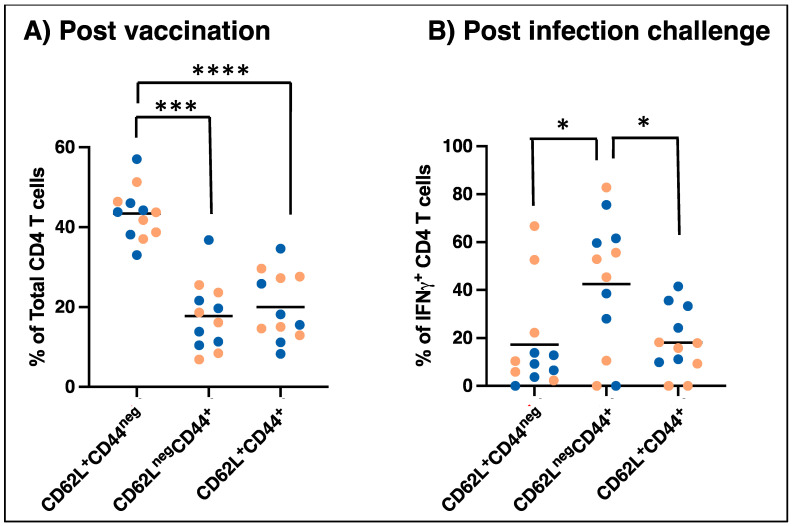
Human monocytic ehrlichiosis, an emerging tick-borne disease, is caused by Ehrlichia chaffeensis. Infections with the pathogen are also common in the canine host. Our previous studies demonstrated that functional disruption within the E. chaffeensis phage head-to-tail connector protein gene results in bacterial attenuation, creating a modified live attenuated vaccine (MLAV). The MLAV confers protective immunity against intravenous and tick transmission challenges one month following vaccination. In this study, we evaluated the duration of MLAV protection. Dogs vaccinated with the MLAV were challenged with wild-type E. chaffeensis via intravenous infection at 4-, 8-, and 12-months post-vaccination. Immunized dogs rapidly cleared the wild-type pathogen infection and tested positive for bacteremia less frequently than unvaccinated controls. While immune responses varied among dogs, vaccinees consistently mounted IgG and CD4+ T-cell responses specific to E. chaffeensis throughout the assessment period. Our findings demonstrate that MLAV-mediated immune protection persists for at least one year against wild-type bacterial infection, marking a major advancement in combating this serious tick-borne disease. The data presented here serve as the foundation for further studies, elucidating the molecular mechanisms underlying virulence and vaccine development and aiding in preventing the diseases caused by E. chaffeensis and other tick-borne rickettsial pathogens.

Keywords: Ehrlichia chaffeensis; immune protection; long-lasting immunity; modified live vaccine; monocytic ehrlichiosis; rickettsiales; tick-borne disease.


Conflict of interest statement

The authors declare no conflicts of interest. The funders had no role in the design of the study; in the collection, analyses, or interpretation of data; in the writing of the manuscript; or in the decision to publish the results.

Figures

Figure 1
Figure 1
MLAV vaccination induced a significant drop in systemic infection. We assessed the differences in the percent positives of PCR detection of infection in the three vaccinated groups and compared them with unvaccinated groups for statistical significance. The one-way ANOVA with multiple comparisons identified significant differences between Groups 1, 2, and 3 compared to Group 4 (unvaccinated) with p < 0.0001 (****) for Groups 1 and 3 and p = 0.0001 (***) for Group 2. There was no significant difference observed when we compared among the three vaccinated groups: Groups 1, 2, and 3.
Figure 2
Figure 2
Antibody response of dogs after MLAV vaccination and after wild-type E. chaffeensis infection challenge. E. chaffeensis-specific IgG was measured in plasma at the indicated time points following vaccination and wild-type challenge for dogs immunized 4, 8, and 12 months prior to infection (groups 1, 2, and 3, respectively). Group 4 dogs were unvaccinated and served as infection controls. IgG responses were analyzed by coating ELISA plates with E. chaffeensis antigens generated from cultures that were either grown in DH82 canine macrophage cell line ((A) left panels) or ISE6 tick cells ((B) right panels). The lines represent the dogs in each group. Females and males were identified with pink and blue lines, respectively. Dogs positive for E. chaffeensis bacteremia at least once following the infection challenge were indicated by arrows in the post-infection challenge images in panel (A).
Figure 3
Figure 3
IFNγ ELISpot responses of dogs following MLAV vaccination and after wild-type infection challenge. PBMCs were isolated at the indicated times following vaccination for the three vaccinated groups ((left) panel) and after infection challenge for all four groups ((right) panel) and plated at a concentration of 4 × 10⁵ cells/well on prepared ELISPOT plates. Cells were stimulated in duplicate wells with 10 μg/mL E. chaffeensis host cell-free lysate. Positive control wells were stimulated with concanavalin A. Negative control wells were stimulated with media only. After overnight culture, ELISPOT plates were developed, and spot-forming units were enumerated using an ELISPOT reader. The lines represent the dogs in each group. Females and males were identified with pink and blue lines, respectively. Dogs positive for E. chaffeensis bacteremia at least once following the infection challenge were indicated by arrows.
Figure 4
Figure 4
IFNγ ELISA responses of dogs following MLAV vaccination (panel (A)) and 4, 8, or 12 months later by intravenous challenge with wild-type E. chaffeensis (panel (B)). PBMCs were isolated and plated, as in Figure 3. Cells were stimulated, as shown in Figure 3, except that the cultures were maintained for 72 h. Culture supernatants were analyzed by commercial sandwich ELISA for IFNγ [p = 0.029 (*) for vaccinated compared to unvaccinated dogs] (Pink and blue bullets refer to female and male dogs in the study, respectively).
Figure 5
Figure 5
Memory CD4 T cell populations in dogs following MLAV vaccination (A) and followed 4, 8, or 12 months later by wild-type E. chaffeensis IV challenge (B). PBMCs were isolated from vaccinated dogs at 8 weeks post-vaccination and stimulated overnight in duplicate wells with 10 μg/mL E. chaffeensis host cell-free lysate, and the assays included positive and controls, as in Figure 3. In the final 5–6 h of culture, Brefeldin A was added to each culture. Cells were then surface stained with antibodies specific to canine CD3, CD4, CD44, and CD62L, and stained intracellularly for IFNγ and used for flow cytometry analysis. Total and E. chaffeensis-specific (gated on IFNγ+) CD4 T cells were analyzed to identify naïve (CD62L+CD44neg), effector memory T cells (CD62LnegCD44+), and central memory T cells (CD62L+CD44+). The dots represent the dogs in each group. Females and males were identified with pink and blue dots, respectively [p = 0.0001 or <0.0001 (*** or ****) for differences between the three naïve, Tcm, and Tem subsets. p = 0.016 (*) for differences between the three IFNg+ naïve, Tcm, and Tem subsets] (Pink and blue bullets refer to female and male dogs in the study, respectively).

Similar articles

Cited by

References

    1. Breitschwerdt E.B., Hegarty B.C., Qurollo B.A., Saito T.B., Maggi R.G., Blanton L.S., Bouyer D.H. Intravascular persistence of Anaplasma platys, Ehrlichia chaffeensis, and Ehrlichia ewingii DNA in the blood of a dog and two family members. Parasit. Vectors. 2014;7:1756–3305. doi: 10.1186/1756-3305-7-298. - DOI - PMC - PubMed
    1. Dumler J.S., Sutker W.L., Walker D.H. Persistent Infection with Ehrlichia chaffeensis. Clin. Infect. Dis. 1993;17:903–905. doi: 10.1093/clinids/17.5.903. - DOI - PubMed
    1. Paddock C.D., Childs J.E. Ehrlichia chaffeensis: A prototypical emerging pathogen. Clin. Microbiol. Rev. 2003;16:37–64. doi: 10.1128/CMR.16.1.37-64.2003. - DOI - PMC - PubMed
    1. Dugan V.G., Little S.E., Stallknecht D.E., Beall A.D. Natural infection of domestic goats with Ehrlichia chaffeensis. J. Clin. Microbiol. 2000;38:448–449. doi: 10.1128/JCM.38.1.448-449.2000. - DOI - PMC - PubMed
    1. Kocan A.A., Levesque G.C., Whitworth L.C., Murphy G.L., Ewing S.A., Barker R.W. Naturally occurring Ehrlichia chaffeensis infection in coyotes from Oklahoma. Emerg. Infect. Dis. 2000;6:477–480. doi: 10.3201/eid0605.000505. - DOI - PMC - PubMed

LinkOut - more resources