. 2024 Aug 13;12(8):914.
doi: 10.3390/vaccines12080914.

Novel Competitive ELISA Utilizing Trimeric Spike Protein of SARS-CoV-2, Could Identify More Than RBD-RBM Specific Neutralizing Antibodies in Hybrid Sera

Affiliations

Novel Competitive ELISA Utilizing Trimeric Spike Protein of SARS-CoV-2, Could Identify More Than RBD-RBM Specific Neutralizing Antibodies in Hybrid Sera

Petros Eliadis et al. Vaccines (Basel). .

Abstract

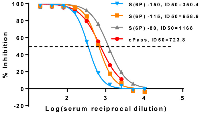
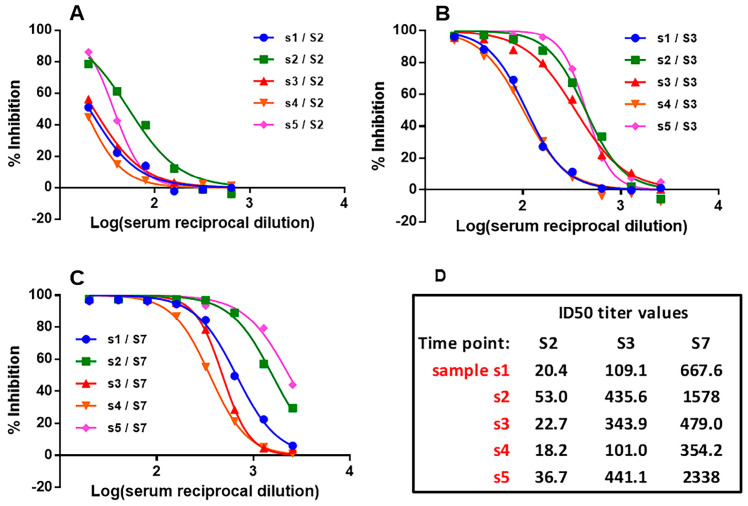
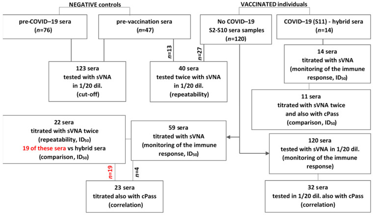
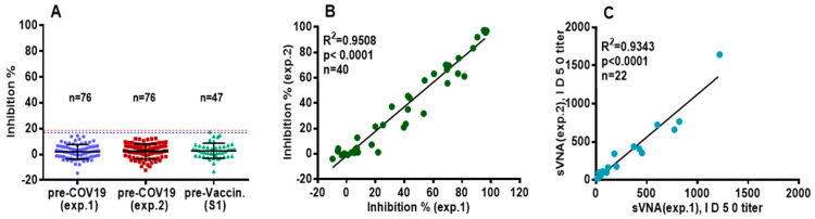
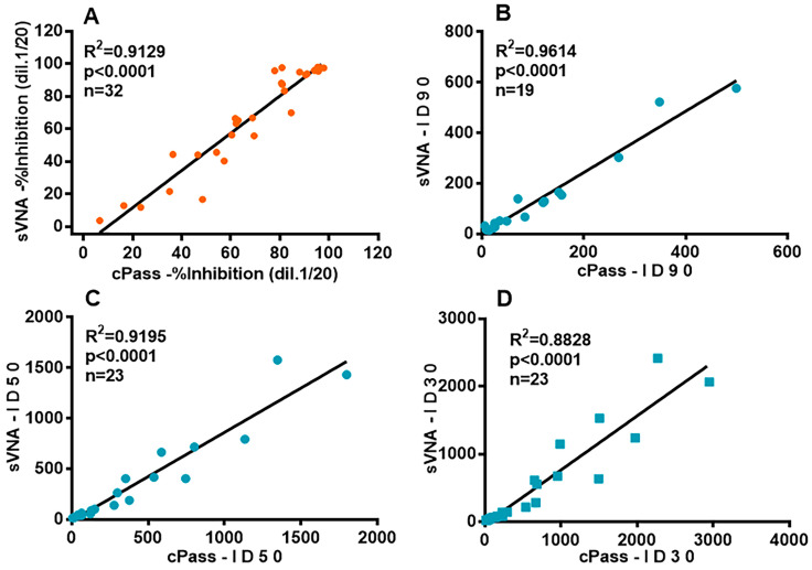
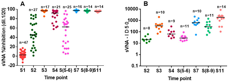
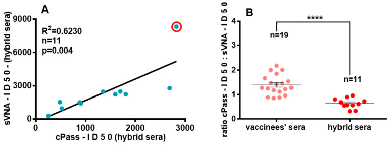
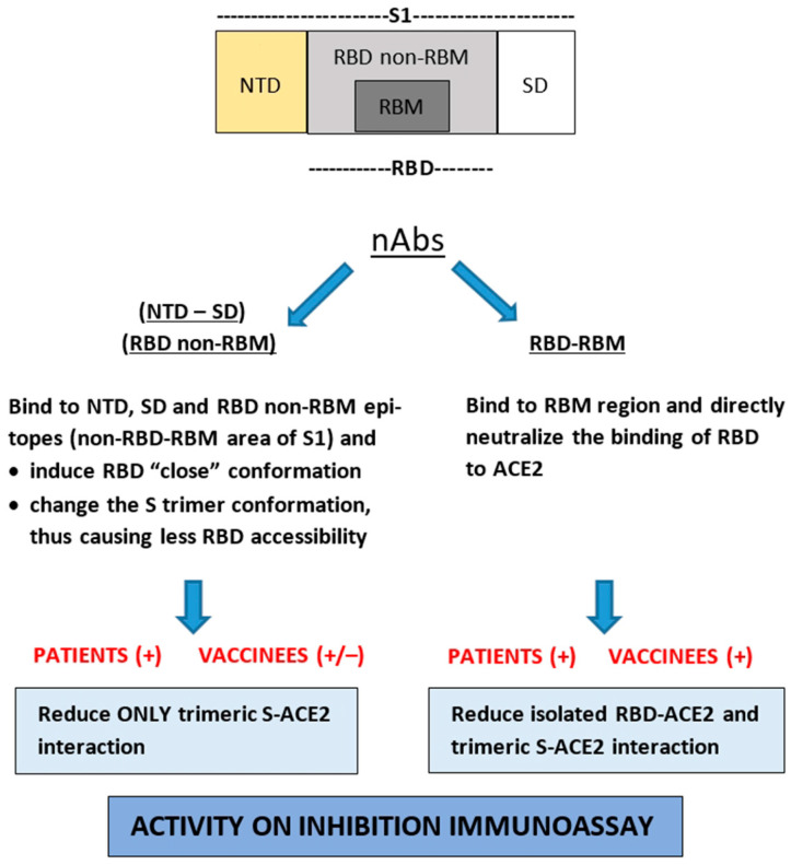
Since the initiation of the COVID-19 pandemic, there has been a need for the development of diagnostic methods to determine the factors implicated in mounting an immune response against the virus. The most promising indicator has been suggested to be neutralizing antibodies (nAbs), which mainly block the interaction between the Spike protein (S) of SARS-CoV-2 and the host entry receptor ACE2. In this study, we aimed to develop and optimize conditions of a competitive ELISA to measure serum neutralizing titer, using a recombinant trimeric Spike protein modified to have six additional proline residues (S(6P)-HexaPro) and h-ACE2. The results of our surrogate Virus Neutralizing Assay (sVNA) were compared against the commercial sVNT (cPass, Nanjing GenScript Biotech Co., Nanjing City, China), using serially diluted sera from vaccinees, and a high correlation of ID50-90 titer values was observed between the two assays. Interestingly, when we tested and compared the neutralizing activity of sera from eleven fully vaccinated individuals who subsequently contracted COVID-19 (hybrid sera), we recorded a moderate correlation between the two assays, while higher sera neutralizing titers were measured with sVNA. Our data indicated that the sVNA, as a more biologically relevant model assay that paired the trimeric S(6P) with ACE2, instead of the isolated RBD-ACE2 pairing cPass test, could identify nAbs other than the RBD-RBM specific ones.

Keywords: SARS-CoV-2; non-RBD-RBM neutralizing antibodies (nAbs); surrogate viral neutralization assay (sVNA); trimeric S(6P)-HexaPro.


Conflict of interest statement

The authors declare no conflicts of interest. The funders had no role in the design of the study; in the collection, analyses, or interpretation of data; in the writing of the manuscript, or in the decision to publish the results.

Figures

Figure 1
Figure 1
Graphical illustration of the developed surrogate Virus Neutralization Assay (sVNA). (I) Binding of SARS-CoV-2 trimeric spike S6(P)-StrepTag to hACE2, no nAbs; (II) (A) Direct blocking of S6(P)-StrepTag from binding to hACE2 by anti-RBM nAbs, (B) Indirect inhibition of S(6P)-ACE2 interaction by anti-RBD non-RBM nAbs, anti-NDT nAbs and anti-SD nAbs.
Figure 2
Figure 2
Titration curves of sVNA and cPass. Evaluation of the effect different concentrations of S(6P)-StrepTag have on the dose-dependent percent inhibition curves of a serum sample (S8). The red line represents the curve created from this sample using the commercial cPass test, that is similar to the curve (orange line) created by 115 ng/mL final concentration of S(6P) when using the sVNA. The experiment was repeated with three different sera in duplicates each time. A representative experiment is shown. The dots represent the mean values from these two replicates. The dotted line indicates the ID50* inhibition level. * The Inhibitory Dilution (ID) at which the serum was able to reduce the % inhibitory effects by 50% of the maximal S(6P)-ACE2 interaction, observed in the absence of serum, was designated as the sVNA-ID50 value.
Figure 3
Figure 3
sVNA titration curves. Dose dependent inhibition of the interaction between immobilized ACE2 and soluble S(6P) in the presence of 5 representative sera (s): (A) S2 (21d after 1st dose of the vaccine), (B) S3 (21d after 2nd dose of the vaccine), (C) samples S7 (21d after 3rd dose of the vaccine). (D) ID50 values were determined as described in the method section, and are presented herein.
Figure 4
Figure 4
Recruitment, testing of sera and follow-up. In this study sera from non-vaccinated and vaccinated individuals (following administration of the first, second, and third dose of the BNT1−62b2 vaccine), as well as fully vaccinated patients infected with COVID−19 (hybrid sera) were used.
Figure 5
Figure 5
Testing sera with sVNAssay. (A) Sera of pre-COVID-19 (n = 76) at the final dilution of 1:20, were tested twice on two different experiments, and sera of pre-vaccinated individuals (S1, n = 47) at the final dilution of 1:20 were tested once. The horizontal lines indicate the Mean values ± SD. The dotted lines (3 × SD) represent the cutoff at ~20% Inhibition. (B) Sera from vaccinated (n = 27) and pre-vaccinated (n = 13) subjects were tested, at dilution 1:20, on two different experiments for their neutralizing ability and a correlation of their inhibition values was performed. (C) Twenty-two vaccinees’ sera were tested, in serial dilutions, on two different experiments and a correlation of their ID50 values was performed.
Figure 6
Figure 6
Correlation analysis for vaccinees’ sera with different levels of nAbs by sVNA and cPass assay. (A) Thirty-two sera from vaccinated individuals were tested in duplicate by sVNA and cPass, at a final serum dilution of 1:20. Mean values are shown. (BD) The data presented are the ID90, ID50 and ID30 titer values, respectively, from twenty-three vaccinees’ serially diluted sera (with inhibition values ranging from 50% to 100% at 1:20 final dilution), tested with sVNA (the 19 sera titrated twice in two different experiments and the two ID50–90–30 titer values were averaged (see Figure 5C)) or cPass, with each dilution response tested in duplicate or triplicate. In panel 3B, four of the twenty three ID90 values could not be calculated, because of low nAbs titers.
Figure 7
Figure 7
Testing sera samples derived from vaccinated persons with sVNA. (A) Time-course of neutralizing anti-S(6P) antibodies. Inhibition (%) of sera samples (S1–S11) at the final dilution of 1:20, collected before (S1), throughout the vaccination period (S2–S9) and after COVID-19 disease (S11). The horizontal lines indicate the median values. (B) Fifty-nine vaccinees’ sera collected from twelve individuals (all samples collected after the first vaccine dose (S2) induced >50% inhibition at the final dilution of 1:20) throughout the vaccination period, and fourteen patients’ sera were tested in serial dilutions and their ID50 values are presented. Indication of different samples refers to the status of each serum cohort (Section 2, Table 1).
Figure 8
Figure 8
Testing vaccinated individuals and hybrid sera with sVNA and cPass. (A) Correlation of ID50 titer values of eleven hybrid sera. In the red circle, the “outlier value”. (B) Differences between the ratios of cPass-ID50: sVNA-ID50 titer values from fully vaccinated individuals and hybrid sera (**** p < 0.0001). The whole group of values are normally distributed; a two-sample t-test was used. Averaged mean values ± SEM are shown.
Figure 9
Figure 9
Schematic representation of nAbs targeting different S1 subdomains of SARS-CoV-2 Spike protein [RBD (receptor binding domain, consisting of RBM and non-RBM region), NTD (aminoterminal domain), SD (subdomain-intermediate region)] and their potential effect in the ectodomain S-trimer and isolated RBD interaction with ACE2.

Similar articles

Cited by

References

    1. Beyerstedt S., Casaro E.B., Rangel É.B. COVID-19: Angiotensin-Converting Enzyme 2 (ACE2) Expression and Tissue Susceptibility to SARS-CoV-2 Infection. Eur. J. Clin. Microbiol. Infect. Dis. 2021;40:905–919. doi: 10.1007/s10096-020-04138-6. - DOI - PMC - PubMed
    1. Lan J., Ge J., Yu J., Shan S., Zhou H., Fan S., Zhang Q., Shi X., Wang Q., Zhang L., et al. Structure of the SARS-CoV-2 Spike Receptor-Binding Domain Bound to the ACE2 Receptor. Nature. 2020;581:215–220. doi: 10.1038/s41586-020-2180-5. - DOI - PubMed
    1. Wolf K.A., Kwan J.C., Kamil J.P. Structural Dynamics and Molecular Evolution of the SARS-CoV-2 Spike Protein. MBio. 2022;13:e02030-21. doi: 10.1128/mbio.02030-21. - DOI - PMC - PubMed
    1. Chen Y., Zhao X., Zhou H., Zhu H., Jiang S., Wang P. Broadly Neutralizing Antibodies to SARS-CoV-2 and Other Human Coronaviruses. Nat. Rev. Immunol. 2023;23:189–199. doi: 10.1038/s41577-022-00784-3. - DOI - PMC - PubMed
    1. Ray D., Le L., Andricioaei I. Distant Residues Modulate Conformational Opening in SARS-CoV-2 Spike Protein. Proc. Natl. Acad. Sci. USA. 2021;118:e2100943118. doi: 10.1073/pnas.2100943118. - DOI - PMC - PubMed

LinkOut - more resources