. 2024 Aug 5;17(8):1034.
doi: 10.3390/ph17081034.

Synthesis and Biochemical Evaluation of Ethanoanthracenes and Related Compounds: Antiproliferative and Pro-Apoptotic Effects in Chronic Lymphocytic Leukemia (CLL)

Affiliations

Synthesis and Biochemical Evaluation of Ethanoanthracenes and Related Compounds: Antiproliferative and Pro-Apoptotic Effects in Chronic Lymphocytic Leukemia (CLL)

James P McKeown et al. Pharmaceuticals (Basel). .

Abstract

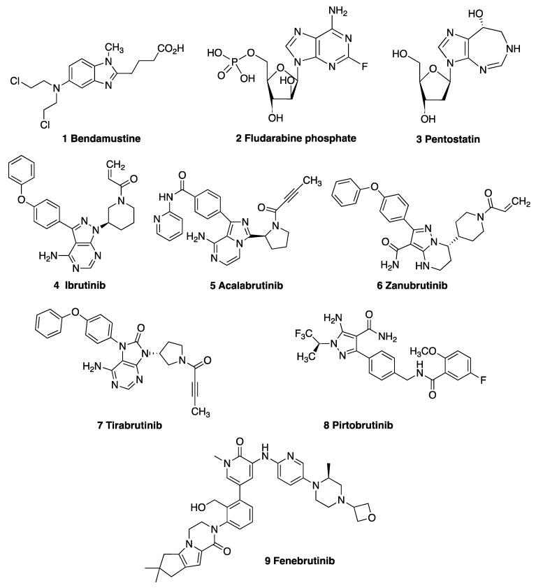
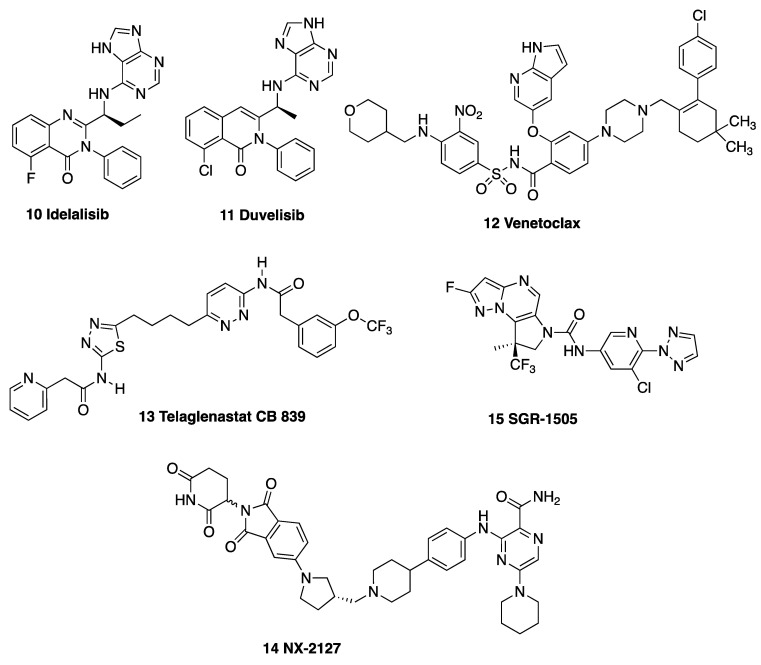
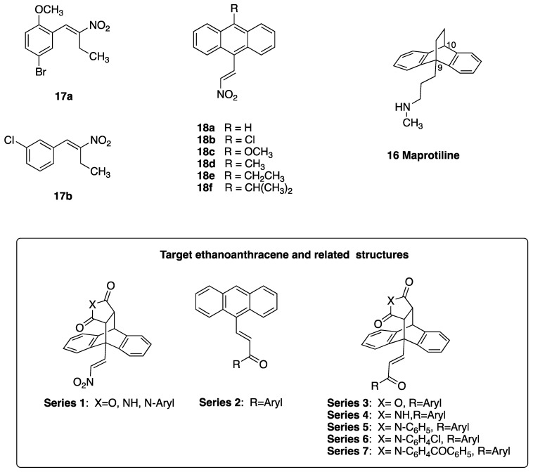
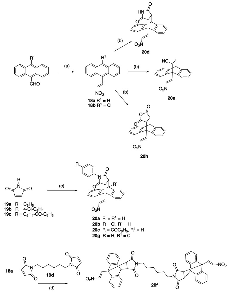
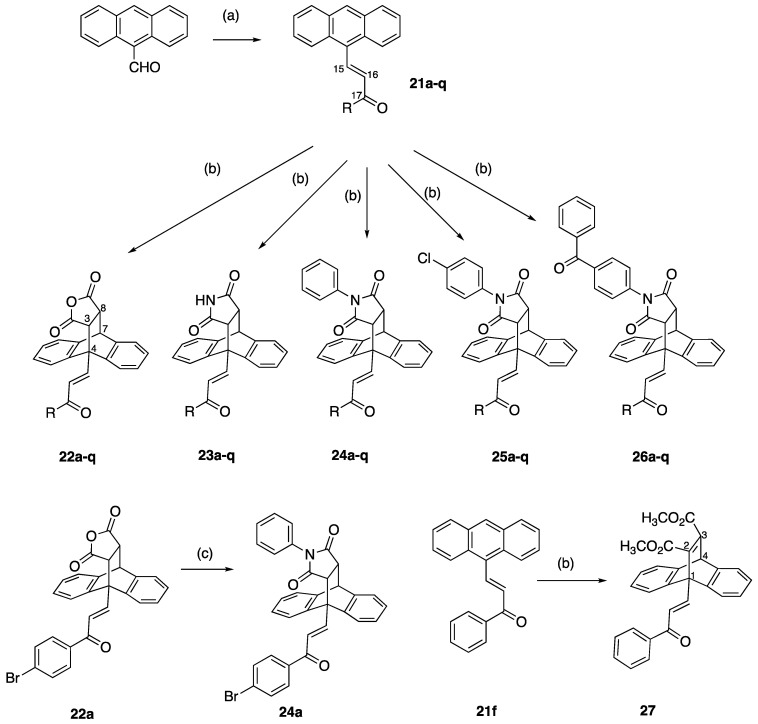
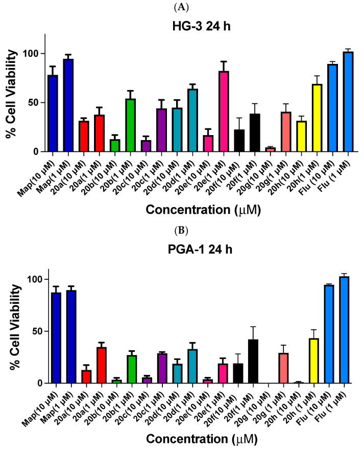
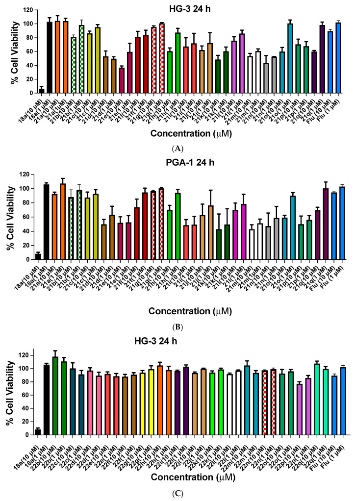
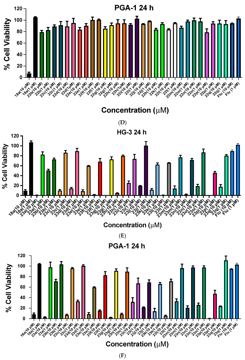
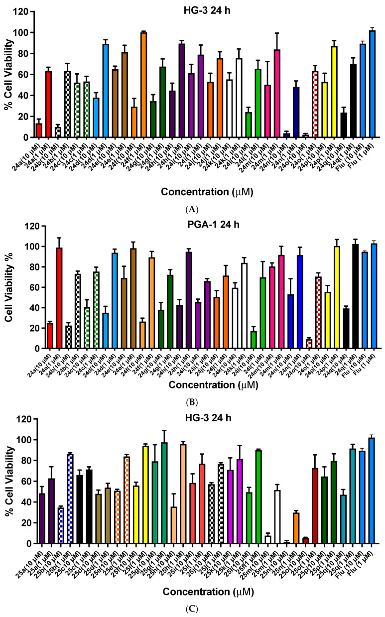
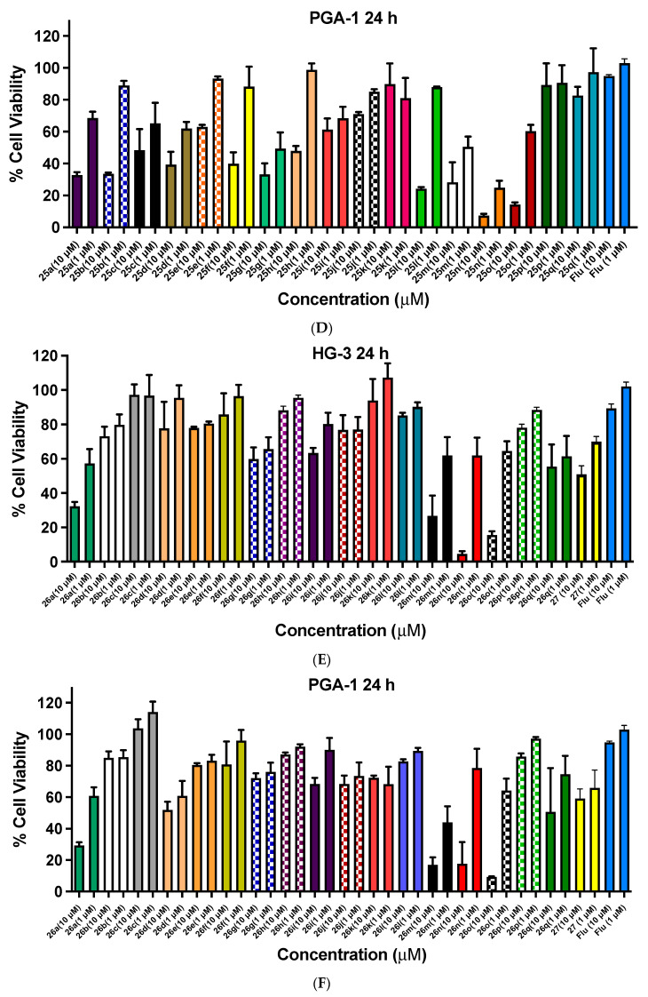
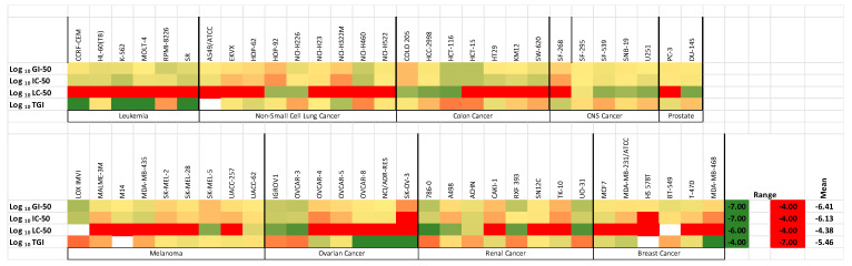
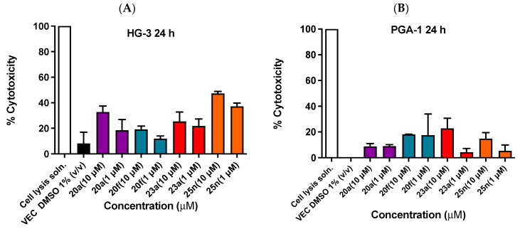
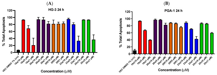
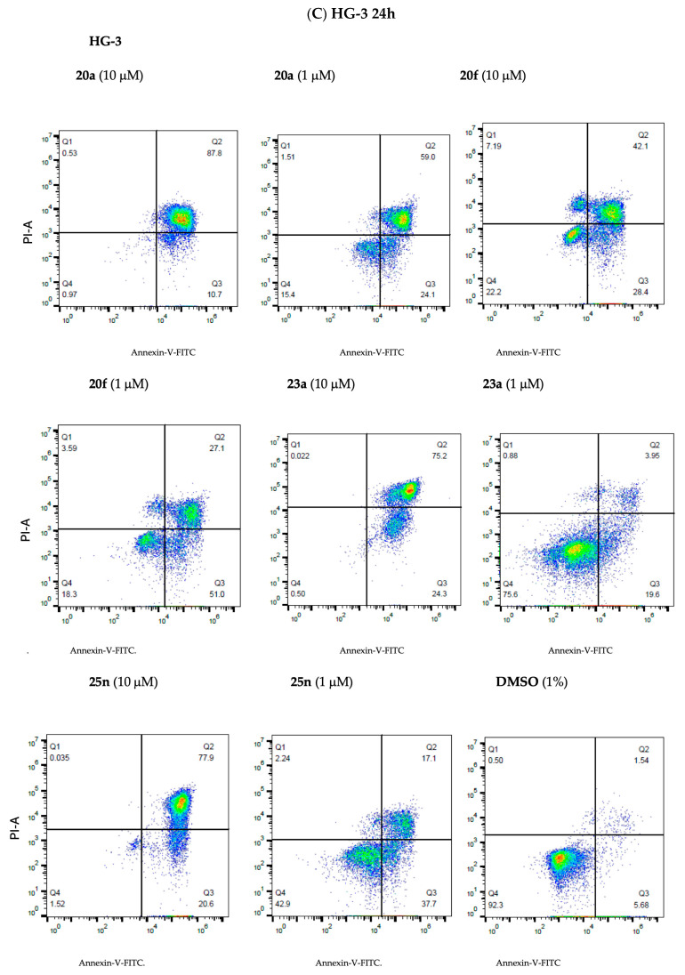
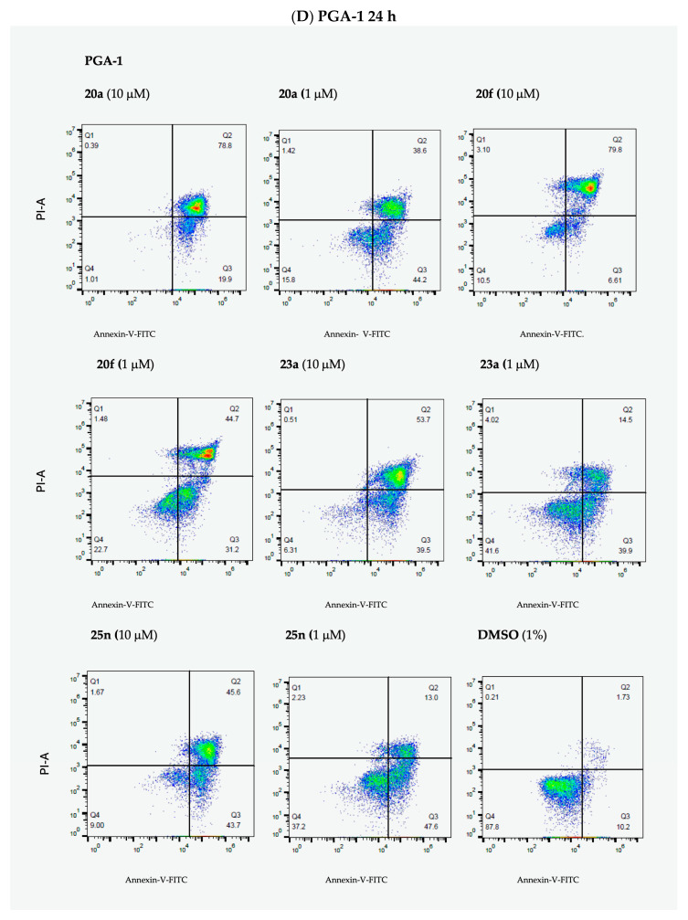
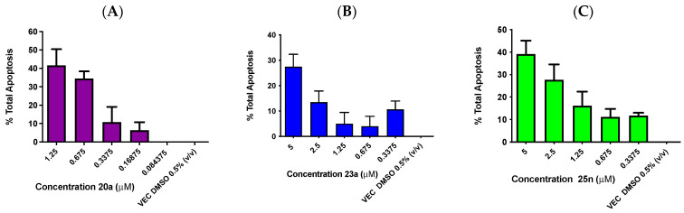
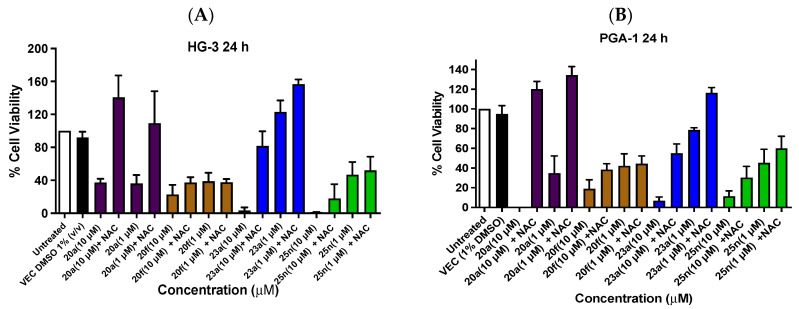
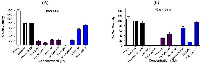
Chronic lymphocytic leukemia (CLL) is a malignancy of mature B cells, and it is the most frequent form of leukemia diagnosed in Western countries. It is characterized by the proliferation and accumulation of neoplastic B lymphocytes in the blood, lymph nodes, bone marrow and spleen. We report the synthesis and antiproliferative effects of a series of novel ethanoanthracene compounds in CLL cell lines. Structural modifications were achieved via the Diels-Alder reaction of 9-(2-nitrovinyl)anthracene and 3-(anthracen-9-yl)-1-arylprop-2-en-1-ones (anthracene chalcones) with dienophiles, including maleic anhydride and N-substituted maleimides, to afford a series of 9-(E)-(2-nitrovinyl)-9,10-dihydro-9,10-[3,4]epipyrroloanthracene-12,14-diones, 9-(E)-3-oxo-3-phenylprop-1-en-1-yl)-9,10-dihydro-9,10-[3,4]epipyrroloanthracene-12,14-diones and related compounds. Single-crystal X-ray analysis confirmed the structures of the novel ethanoanthracenes 23f, 23h, 24a, 24g, 25f and 27. The products were evaluated in HG-3 and PGA-1 CLL cell lines (representative of poor and good patient prognosis, respectively). The most potent compounds were identified as 20a, 20f, 23a and 25n with IC50 values in the ranges of 0.17-2.69 µM (HG-3) and 0.35-1.97 µM (PGA-1). The pro-apoptotic effects of the potent compounds 20a, 20f, 23a and 25n were demonstrated in CLL cell lines HG-3 (82-95%) and PGA-1 (87-97%) at 10 µM, with low toxicity (12-16%) observed in healthy-donor peripheral blood mononuclear cells (PBMCs) at concentrations representative of the compounds IC50 values for both the HG-3 and PGA-1 CLL cell lines. The antiproliferative effect of the selected compounds, 20a, 20f, 23a and 25n, was mediated through ROS flux with a marked increase in cell viability upon pretreatment with the antioxidant NAC. 25n also demonstrated sub-micromolar activity in the NCI 60 cancer cell line panel, with a mean GI50 value of 0.245 µM. This ethanoanthracene series of compounds offers potential for the further development of lead structures as novel chemotherapeutics to target CLL.

Keywords: B cell; antiproliferative; chalcone; chronic lymphocytic leukemia (CLL); ethanoanthracene; pro-apoptotic.


Conflict of interest statement

The authors declare no conflicts of interest.

Figures

Figure 1
Figure 1
Drugs used in the treatment of CLL: alkylating agents 1 bendamustine, 2 fludarabine phosphate and 3 pentostatin; covalent BTK inhibitors 4 ibrutinib, 5 acalabrutinib, 6 zanubrutinib and 7 tirabrutinib; and non-covalent BTK inhibitors 8 pirtobrutinib and 9 fenebrutinib.
Figure 2
Figure 2
Drugs targeting CLL: PI3Kδ inhibitor idelalasilib 10; PI3Kδ and PI3Kγ inhibitor duvelisib 11; Bcl-2 inhibitor venetoclax 12; glutaminase inhibitor telaglenastat CB 839 13; and dual BTK degrader NX-2127 14 and MALT-1 inhibitor SGR-1505 15.
Figure 3
Figure 3
Nitrostyrenes 17a, 17b, nitrovinylanthracenes 18a–e and maprotiline 16; target ethanoanthracene structures, Series 1–7.
Scheme 1
Scheme 1
Synthesis of Series 1 ethanoanthracenes 20a–g reagents and conditions: (a) piperidine acetate, excess nitromethane (CH3NO2), 90 °C, N2, 1.5 h (71–99%); (b) dienophile (maleic anhydride) for 20h, maleimide for 20d, NCHC=CH2 for 20e, toluene, 90 °C, 48 h (30–80%); (c) dienophile 19a for N-arylmaleimides 20a and 20g, 19b for 20g, 19c for 20c, toluene, 90 °C, 48 h, (15–51%); and (d) toluene, 90 °C, 48 h (10%).
Scheme 2
Scheme 2
Synthesis of Series 2 anthracene chalcones 21a–q and Series 3–7 ethanoanthracenes 22a–q, 23a–q, 24a–q, 25a–q, 26a–q and 27 (see Table 1 for substituents and yields). Reagents and conditions: (a) Appropriate aryl methyl ketone, EtOH, NaOH, 20 °C, 24 h. (b) Appropriate anthracene chalcone 21a–q, dieneophile (maleic anhydride for 22a–q, maleimide for 23a–q, N-phenylmaleimide for 24a–q, N-(4-chlorophenyl)maleimide for 25a–q, N-(4-benzoylphenyl)maleimide for 26a–q, dimethyl acetylenedicarboxylate for 27), toluene, 90 °C, 48 h. (c) Aniline, acetic acid 120 °C, 2–3 h (72%).
Figure 4
Figure 4
Stability study for compounds 21a, 21i, 22h, 23a, 23g, 23n, 24a, 24h, 26a and 26n at pH 4.0, pH 7.5 and pH 9.0 over 24 h.
Figure 5
Figure 5
Cell viability data for ethanoanthracenes 20a–e, 20g, 20h and 20f. Cell viability data for (E)-9-(2-Nitrovinyl)-9,10,11,15-tetrahydro-9,10-[3,4]epipyrroloanthracene-12,14-diones 20a–e, 20g and 20h and the related dimer 20f in CLL: (A) HG-3 cells (1 and 10 µM) and (B) PGA-1 cells (1 and 10 µM). The cell proliferation of HG-3 and PGA-1 cells was determined with an alamarBlue assay. Compound concentrations of either 1 µM or 10 µM for 24 h were used to treat the cells (in triplicate) with control wells containing vehicle DMSO (1% v/v). Map = maprotiline; Flu = fludarabine. The mean value for three experiments is shown.
Figure 6
Figure 6
Cell viability data for chalcones 21a–q and ethanoanthracenes 22a–22q and 23a–23q in CLL cell lines HG-3 and PGA-1. Cell viability data for chalcones 21a–q (A,B), maleic anhydride ethanoanthracene adducts 22a–22q (C,D) and maleimide ethanoanthracene adducts 23a–23q (E,F) in CLL cell lines: the cell proliferation of HG-3 and PGA-1 cells was determined with an alamarBlue assay. Compound concentrations of either 1 µM or 10 µM for 24 h were used to treat the cells (in triplicate) with control wells containing vehicle DMSO (1% v/v). Flu = fludarabine. The mean value for three independent experiments is shown.
Figure 6
Figure 6
Cell viability data for chalcones 21a–q and ethanoanthracenes 22a–22q and 23a–23q in CLL cell lines HG-3 and PGA-1. Cell viability data for chalcones 21a–q (A,B), maleic anhydride ethanoanthracene adducts 22a–22q (C,D) and maleimide ethanoanthracene adducts 23a–23q (E,F) in CLL cell lines: the cell proliferation of HG-3 and PGA-1 cells was determined with an alamarBlue assay. Compound concentrations of either 1 µM or 10 µM for 24 h were used to treat the cells (in triplicate) with control wells containing vehicle DMSO (1% v/v). Flu = fludarabine. The mean value for three independent experiments is shown.
Figure 7
Figure 7
Cell viability data for ethanoanthracenes 24a–q, 25a–q, 26a–q and 27 in CLL cell lines HG-3 and PGA-1. Cell viability data for N-phenylmaleimide-substituted ethanoanthracenes (24a–q, Panels A,B), N-(4-chlorophenyl)maleimide-substituted ethanoanthracenes (25a–q, Panels C and D) and N-(4-benzoylphenyl)maleimide-substituted ethanoanthracenes (26a–q, Panels E,F) and 27 (E,F) were determined in CLL cells HG-3 cells (1 and 10 µM) and PGA-1 cells (1 and 10 µM). The cell proliferation of HG-3 and PGA-1 cells was determined with an alamarBlue assay. Compound concentrations of either 1 µM or 10 µM for 24 h were used to treat the cells (in triplicate) with control wells containing vehicle DMSO (1% v/v). Flu = fludarabine. The mean value for three independent experiments is shown.
Figure 7
Figure 7
Cell viability data for ethanoanthracenes 24a–q, 25a–q, 26a–q and 27 in CLL cell lines HG-3 and PGA-1. Cell viability data for N-phenylmaleimide-substituted ethanoanthracenes (24a–q, Panels A,B), N-(4-chlorophenyl)maleimide-substituted ethanoanthracenes (25a–q, Panels C and D) and N-(4-benzoylphenyl)maleimide-substituted ethanoanthracenes (26a–q, Panels E,F) and 27 (E,F) were determined in CLL cells HG-3 cells (1 and 10 µM) and PGA-1 cells (1 and 10 µM). The cell proliferation of HG-3 and PGA-1 cells was determined with an alamarBlue assay. Compound concentrations of either 1 µM or 10 µM for 24 h were used to treat the cells (in triplicate) with control wells containing vehicle DMSO (1% v/v). Flu = fludarabine. The mean value for three independent experiments is shown.
Figure 8
Figure 8
Heatmap for compound 25n across cell lines in the NCI-60 screen.
Figure 9
Figure 9
LDH assay for cytotoxicity of compounds 20a, 20f, 23a and 25n in the HG-3 (Panel A) and PGA-1 (Panel B) cell lines.
Figure 10
Figure 10
Ethanoanthracene nitrostyrene compounds 18a, 20a, 20f, 23a and 25n induce apoptosis in HG-3 and PGA-1 CLL cells. Compounds 18a, 20a, 20f, 23a and 25n potently induce apoptosis in HG-3 and PGA-1 cell lines (Annexin V/PI FACS). HG-3 and PGA-1 leukemia cells were treated with 18a, 20a, 20f, 23a and 25n (10 µM, 5 µM and 1 µM) and a control vehicle [(1% DMSO (v/v))] at 48 h for panels A and B, respectively. The % of apoptotic cells was determined by staining with Annexin V-FITC and PI (Panels C and D show compounds 20a, 20f, 23a and 25n at 1- and 10-µM concentrations). The lower left quadrant cells are negative for both Annexin V-FITC and PI, and the upper left shows PI cells that are necrotic. The lower right quadrant shows Annexin-positive cells in the early apoptotic stage, and the upper right shows both Annexin- and PI-positive cells in the late apoptosis stage. The experiment was replicated on three independent days.
Figure 10
Figure 10
Ethanoanthracene nitrostyrene compounds 18a, 20a, 20f, 23a and 25n induce apoptosis in HG-3 and PGA-1 CLL cells. Compounds 18a, 20a, 20f, 23a and 25n potently induce apoptosis in HG-3 and PGA-1 cell lines (Annexin V/PI FACS). HG-3 and PGA-1 leukemia cells were treated with 18a, 20a, 20f, 23a and 25n (10 µM, 5 µM and 1 µM) and a control vehicle [(1% DMSO (v/v))] at 48 h for panels A and B, respectively. The % of apoptotic cells was determined by staining with Annexin V-FITC and PI (Panels C and D show compounds 20a, 20f, 23a and 25n at 1- and 10-µM concentrations). The lower left quadrant cells are negative for both Annexin V-FITC and PI, and the upper left shows PI cells that are necrotic. The lower right quadrant shows Annexin-positive cells in the early apoptotic stage, and the upper right shows both Annexin- and PI-positive cells in the late apoptosis stage. The experiment was replicated on three independent days.
Figure 11
Figure 11
Percentage of total apoptosis observed upon treatment of isolated donor PBMCs with compounds 20a, 23a and 25n. Ethanoanthracene compounds 20a, 23a and 25n induced apoptosis upon the treatment of isolated human-donor peripheral blood mononuclear cells (PBMCs). Isolated donor PBMCs were treated with compound 20a (Panel A) at 1.25 µM, 0.68 µM, 0.34 µM, 0.17 µM and 0.08 µM concentrations, compounds 23a (Panel B) and 25n (Panel C) at 5 µM, 2.5 µM, 1.25 µM, 0.68 µM and 0.34 µM concentrations, and all were normalized against a control vehicle (0.5% DMSO (v/v)) at 48 h. The % of apoptotic cells was determined via staining with Annexin V-FITC and PI. The experiment was performed individually and replicated on three independent days.
Figure 12
Figure 12
Effect of antioxidant pre-treatment (N-acetylcysteine, NAC) on the viability of HG-3 and PGA-1 CLL cells treated with compounds 20a, 20f, 23a and 25n. The cell viability of HG-3 and PGA-1 cells was determined with an alamarBlue assay (seeding density: 2 × 10⁵ cells/mL per well for 96-well plates). Compound concentrations of either 1 µM or 10 µM for 24 h were used to treat the HG-3 and PGA-1 CLL cells (in triplicate) with control wells containing vehicle DMSO (1% v/v). The cells were pre-treated with NAC (2 µL, 5 mM) for 1 h, (Panel A,B) and protected from light before then being treated with the compound. The mean value for three independent experiments is shown.
Figure 13
Figure 13
Effect of pre-treatment with caspase inhibitor Z-VAD-FMK on HG-3 and PGA-1 cell viability for compounds 20a and 23a. Cell viability analysis (24 h) for inhibitor studies of compounds 20a and 23a in HG-3 (Panel A) and PGA-1 (Panel B) CLL cell lines: the HG-3 and PGA-1 CLL cells (2 × 10⁵ cells/mL) were pre-treated at 37 °C with 40 µM of caspase inhibitor (CI) (Z-VAD-FMK) for 4 h prior to compound treatment at 1 µM and 10 µM for 24 h. The cell proliferation of HG-3 and PGA-1 cells was determined with an alamarBlue assay (CI = caspase inhibitor, n = 2).
Figure 14
Figure 14
Cumulative probability scores for compounds 20a, 20b, 20d–20f, 23a, 23c, 23f–23i, 23k, 23l, 23n, 23p, 24f, 24l, 25n and 26n for the 30 targets most strongly indicated via STP.
Figure 15
Figure 15
Standardized cumulative probability scores of target groups indicated via STP for the tested compounds 20a, 20b, 20d–20f, 23a, 23c, 23f–23i, 23k, 23l, 23n, 23p, 24f, 24l, 25n and 26n compared to Maprotiline. Scores were standardized using z-scores (i.e., differences in standard deviations from their mean).

Similar articles

References

    1. Cancer Stat Facts: Leukemia—Chronic Lymphocytic Leukemia (CLL) National Cancer Institute. [(accessed on 26 April 2024)];2022 Available online: https://seer.cancer.gov/statfacts/html/clyl.html.
    1. Cronin K.A., Scott S., Firth A.U., Sung H., Henley S.J., Sherman R.L., Siegel R.L., Anderson R.N., Kohler B.A., Benard V.B., et al. Annual report to the nation on the status of cancer, part 1: National cancer statistics. Cancer. 2022;128:4251–4284. doi: 10.1002/cncr.34479. - DOI - PMC - PubMed
    1. Waldron C., O’Brien D., Brophy S., Perera K., Crotty G.M., Dunlea E., Walsh A., Connolly M., Clifford R., O’Leary H., et al. Epidemiology of chronic lymphocytic leukaemia in an Irish subpopulation with total case ascertainment: An additional tool for health economic planning. Br. J. Haematol. 2022;196:e47–e49. doi: 10.1111/bjh.17929. - DOI - PubMed
    1. Hallek M. Chronic lymphocytic leukemia: 2020 update on diagnosis, risk stratification and treatment. Am. J. Hematol. 2019;94:1266–1287. doi: 10.1002/ajh.25595. - DOI - PubMed
    1. Shadman M. Diagnosis and treatment of chronic lymphocytic leukemia: A review. JAMA. 2023;329:918–932. doi: 10.1001/jama.2023.1946. - DOI - PubMed

LinkOut - more resources