Miamiensis avidus, a Novel Scuticociliate Pathogen Isolated and Identified from Cultured Large Yellow Croaker (Larimichthys crocea)
- PMID: 39204219
- PMCID: PMC11357409
- DOI: 10.3390/pathogens13080618
Miamiensis avidus, a Novel Scuticociliate Pathogen Isolated and Identified from Cultured Large Yellow Croaker (Larimichthys crocea)
Abstract
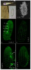
Scuticociliates are recognized as the causative agents of mass mortalities in certain cultured marine fishes, resulting in enormous economic losses. This study aimed to investigate a fatal infection caused by scuticociliates in farmed large yellow croaker (Larimichthys crocea) in Fujian province, China. Microscopic examinations of focal organs, including the brain, eyes, gills, and skin, revealed the presence of parasites. Active masses of scuticociliates were observed in these organs, and the ciliates were subsequently isolated and maintained in vitro. An immersion challenge experiment revealed that L. crocea experienced cumulative mortalities reaching 73% within 7 d upon exposure to 1.0 × 10⁴ ciliates mL-1. Based on the microscopic and PCR testing of infected fishes, the brain was comprehensively inferred as the main infection organ for the isolated strain. Microscopic and submicroscopic observations of the isolated scuticociliate, coupled with cortical ciliature patterns revealed through α-tubulin indirect immunofluorescence techniques, identified these scuticociliates as Miamiensis avidus. The sequencing of two genetic markers (small subunit ribosomal RNA, SSU rRNA and cytochrome c oxidase subunit I, COI) further confirmed that the isolated strains exhibited the highest sequence similarity to most M. avidus sequences in GenBank. However, significant differences in SSU sequences compared to the M. avidus strain Ma/2, and the lack of published COI and ITS (internal transcribed spacer) sequences for Ma/2, indicate the need for further molecular data to resolve whether there are potential cryptic species within the M. avidus complex.
Keywords: L. crocea; Miamiensis avidus; Scuticociliatosis; molecular identification; α-tubulin indirect immunofluorescence.
Conflict of interest statement
The authors declare no conflicts of interest. The funders had no role in the design of the study; in the collection, analyses, or interpretation of data; in the writing of the manuscript; or in the decision to publish the results.
Figures


References
-
- Bureau of Fisheries of Ministry of Agriculture and Rural Affairs. National Fisheries Technology Extension Center & China Society of Fisheries . China Fishery Statistical Yearbook 2023. China Agriculture Press; Beijing, China: 2023. p. 26.
-
- Kong S., Ke Q., Chen L., Zhou Z., Pu F., Zhao J., Bai H., Peng W., Xu P. Constructing a High-Density Genetic Linkage Map for Large Yellow Croaker (Larimichthys crocea) and Mapping Resistance Trait Against Ciliate Parasite Cryptocaryon irritans. Mar. Biotechnol. 2019;21:262–275. doi: 10.1007/s10126-019-09878-x. - DOI - PubMed
-
- Zhang S. Diagnosis and Treatment of a Ciliate Endoparasific in the Large YellowCroaker (Pseudosciaena crocea) J. Fish. Res. 2011;33:58–61.
-
- Zhang Z.X., Wang Z.Y., Fang M., Ye K., Tang X., Zhang D.L. Genome-wide association analysis on host resistance against the rotten body disease in a naturally infected population of large yellow croaker Larimichthys crocea. Aquaculture. 2021;548:737615. doi: 10.1016/j.aquaculture.2021.737615. - DOI
-
- Chi H., Jiang Q., Pan Y., Lin N. Molecular Identification and Phylogenetic Analysis of a Scuticociliate in Large Yellow Croakers. Fujian J. Anim. Husb. Vet. Med. 2023;45:1–6.
MeSH terms
Grants and funding
- 2019R1027-1/Special Fund for Public-interest Scientific Institutions of Science and Technology Plan Projects of Fujian Province
- AC2017-1/General Projects of Fujian Academy of Agricultural Science
- 2020-1/Scientific Research Innovation Program "Xiyuanjiang River Scholarship" of College of Life Sci-ences, Fujian Normal University.
LinkOut - more resources
Full Text Sources

