. 2024 Jul 27;13(8):629.
doi: 10.3390/pathogens13080629.

Characterisation of European Field Goat Prion Isolates in Ovine PrP Overexpressing Transgenic Mice (Tgshp IX) Reveals Distinct Prion Strains

Affiliations

Characterisation of European Field Goat Prion Isolates in Ovine PrP Overexpressing Transgenic Mice (Tgshp IX) Reveals Distinct Prion Strains

Sonja Ernst et al. Pathogens. .

Abstract

After the detection of bovine spongiform encephalopathy (BSE), and a zoonotic transmissible spongiform encephalopathy (TSE) caused by the pathological prion protein (PrPSc) in two goats, the investigation of goat prions became of greater interest. Therefore, a broad collection of European goat TSE isolates, including atypical scrapie, CH1641 and goat BSE as reference prion strains were biochemically characterised and subsequently inoculated into seven rodent models for further analysis (already published results of this comprehensive study are reviewed here for comparative reasons). We report here the histopathological and immunohistochemical data of this goat TSE panel, obtained after the first passage in Tgshp IX (tg-shARQ) mice, which overexpress the ovine prion protein. In addition to the clear-cut discrimination of all reference prion strains from the classical scrapie (CS) isolates, we were further able to determine three categories of CS strains. The investigation further indicates the occurrence of sub-strains that slightly resemble distant TSE strains, such as BSE or CH1641, reinforcing the theory that CS is not a single strain but a mixture of sub-strains, existing at varying extents in one isolate. This study further proved that Tgshp IX is a potent and reliable tool for the in-depth characterisation of prion strains.

Keywords: BSE; CH1641-like; PrPSc; Tgshp IX; atypical scrapie; bovine spongiform encephalopathy; classical scrapie; goat; immunohistochemistry; strain typing; transgenic mice.


Conflict of interest statement

The authors declare no conflicts of interest.

Figures
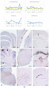
Figure 1
Figure 1
(a). PrPSc profile and cellular PrPSc deposition pattern of reference prion strains. The Tgshp IX PrPSc profile and deposition pattern of the three reference isolates atypical scrapie (AS), caprine BSE (gtBSE) and CH1641 used in the study are presented here. The Tgshp IX model allows for a clear-cut discrimination of all isolates in particular by including the granular and molecular layers of the cerebellum and corpus callosum in the PrPSc profile as well as the overall cellular reaction pattern. NcV = vestibular nuclei of medulla; Tec = tectum of cerebellum; CoS = cortex of the superior colliculus; HTH = hypothalamus; TH = thalamus; HC = hippocampus; Sep = septal nuclei; CbrA = cerebral cortex (at the level of thalamus and hypothalamus); CbrB = cerebral cortex (at the level of the septal nuclei); WM = cerebellar white matter; Dc = white matter in decussation fibres; DpI = internal capsule; CC = corpus callosum; GrL = granular layer of the cerebellar cortex; MoL = molecular layer of the cerebellar cortex; BS = brain stem; CB = cerebellum; MB = midbrain; DE = diencephalon; Cbr = cerebrum; FB = forebrain, CC = corpus callosum. Error bars indicate the standard error of means. (b). Distinct differences between the reference prion strains are seen in cerebellum and diencephalon of representative tg-shARQ (Tgshp IX) mice per group: (AC) atypical scrapie with distinct PrPSc accumulation only in molecular layer of cerebellum and spurious amounts in corpus callosum (arrow); (DF) goat BSE with PrPSc accumulation in granular (arrowhead and inlet) and molecular layer (arrow) of cerebellum as well as distinct subpial reaction pattern in brain stem and cerebellum (E, stars), as well as massive PrPSc depositions in corpus callosum and thalamus, including widespread plaque/plaque-like formations and subpial reaction pattern (F, stars); (GI) CH1641 with a distinct intraneuronal reaction pattern in brain stem associated with fine extracellular PrPSc deposition, and in addition, moderate PrPSc accumulation in corpus callosum. PrP immunohistochemistry mab R145, Bar A, C, D, E, G and I 100 µm, B, H 50 µm.
Figure 2
Figure 2
Mean PrPSc profile summarising the isolates per country with the exception of isolate F16 which is presented separately. Comparative analysis of the mean PrPSc profile of each country. Means from Spain, the Netherlands, France, Greece and Cyprus show a wide homology with peaks at the same brain regions. However, the Italian mean is clearly distinguishable due to its low PrPSc accumulation and the heightened peak at the level of the granular layer of the cerebellum. F16 showed an unique PrPSc profile with an almost complete lack of PrPSc accumulation in CC and WM and a distinct peak in CbrB. NcV = vestibular nuclei of medulla; Tec = tectum of cerebellum; CoS = cortex of the superior colliculus; HTH = hypothalamus; TH = thalamus; HC = hippocampus; Sep = septal nuclei; CbrA = cerebral cortex (at the level of thalamus and hypothalamus); CbrB = cerebral cortex (at the level of the septal nuclei); WM = cerebellar white matter; Dc = white matter in decussation fibres; DpI = internal capsule; CC = corpus callosum; GrL = granular layer of the cerebellar cortex; MoL = molecular layer of the cerebellar cortex. Error bars indicate the standard error of means.
Figure 3
Figure 3
PrPSc profiles of EU mean (CS-1), Italian isolates (CS-2) and F16 (CS-3) compared to reference strains. Country means of Spain, the Netherlands, France, Greece, Cyprus and the UK were summarised to one European mean (EU mean) building category CS-1. The Italian isolates form the category CS-2, and the single F16 isolate is categorised as CS-3. (A) Comparative analysis of the three categories reveals distinct profiles, especially at the level of the corpus callosum (CC) and the granular layer of the cerebellum (GrL). (BD) All categories were compared to reference strains atypical scrapie (AS), caprine BSE (gtBSE) and CH1641. (B) CS-1 was clearly distinguishable from AS and gtBSE, while CH1641 showed more overlapping features to CS-1 in most brain areas investigated, but the lack of PrPSc in the cortex and septum is striking. (C) A clear-cut discrimination from all three reference sequences was possible for CS-2. (D) CS-3 was clearly distinguishable from all references. NcV = vestibular nuclei of medulla; Tec = tectum of cerebellum; CoS = cortex of the superior colliculus; HTH = hypothalamus; TH = thalamus; HC = hippocampus; Sep = septal nuclei; CbrA = cerebral cortex (at the level of thalamus and hypothalamus); CbrB = cerebral cortex (at the level of the septal nuclei); WM = cerebellar white matter; Dc = white matter in decussation fibres; DpI = internal capsule; CC = corpus callosum; GrL = granular layer of the cerebellar cortex; MoL = molecular layer of the cerebellar cortex. Error bars indicate the standard error of means.
Figure 4
Figure 4
Distinct differences between classical scrapie strains (EU mean, Italy and F16) are visible in cerebellum/brain stem, diencephalon and cerebrum (at level of septum, CbrB) of representative tg-shARQ (Tgshp IX) mice per group: (AC) EU mean (CS-1) showing a variable PrPSc deposition pattern, including a clear involvement of corpus callosum, and single areas even show similarities to CH1641 (i.e., distinct intraneuronal reaction pattern in brain stem; see inlet), but CbrB remain free of PrPSc; (DF) Italian cases (CS-2) with distinct PrPSc accumulation in granular layer of cerebellum (arrows, inlet), variable reaction pattern in diencephalon, but PrPSc is neither to be found in corpus callosum nor CbrB; (GI) F16 (CS-3), no PrPSc accumulation in cerebellum and corpus callosum, but distinct deposition in CbrB (stars, inlet); immunohistochemistry mab R145, Bar 100 µm, inlets 50 µm.

References

    1. Aguzzi A., Baumann F., Bremer J. The prion’s elusive reason for being. Annu. Rev. Neurosci. 2008;31:439–477. doi: 10.1146/annurev.neuro.31.060407.125620. - DOI - PubMed
    1. Rivera N.A., Brandt A.L., Novakofski J.E., Mateus-Pinilla N.E. Chronic Wasting Disease In Cervids: Prevalence, Impact And Management Strategies. Vet. Med. 2019;10:123–139. doi: 10.2147/VMRR.S197404. - DOI - PMC - PubMed
    1. Williams E.S., Young S. Chronic wasting disease of captive mule deer: A spongiform encephalopathy. J. Wildl. Dis. 1980;16:89–98. doi: 10.7589/0090-3558-16.1.89. - DOI - PubMed
    1. Babelhadj B., Di Bari M.A., Pirisinu L., Chiappini B., Gaouar S.B.S., Riccardi G., Marcon S., Agrimi U., Nonno R., Vaccari G. Prion Disease in Dromedary Camels, Algeria. Emerg. Infect. Dis. 2018;24:1029–1036. doi: 10.3201/eid2406.172007. - DOI - PMC - PubMed
    1. Wells G.A., Scott A.C., Johnson C.T., Gunning R.F., Hancock R.D., Jeffrey M., Dawson M., Bradley R. A novel progressive spongiform encephalopathy in cattle. Vet. Rec. 1987;121:419–420. doi: 10.1136/vr.121.18.419. - DOI - PubMed

LinkOut - more resources