Review
. 2024 Aug 7;13(8):668.
doi: 10.3390/pathogens13080668.

Aggregatibacter actinomycetemcomitans Dispersin B: The Quintessential Antibiofilm Enzyme

Affiliations
Review

Aggregatibacter actinomycetemcomitans Dispersin B: The Quintessential Antibiofilm Enzyme

Jeffrey B Kaplan et al. Pathogens. .

Abstract

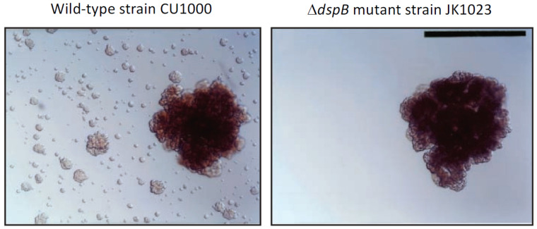
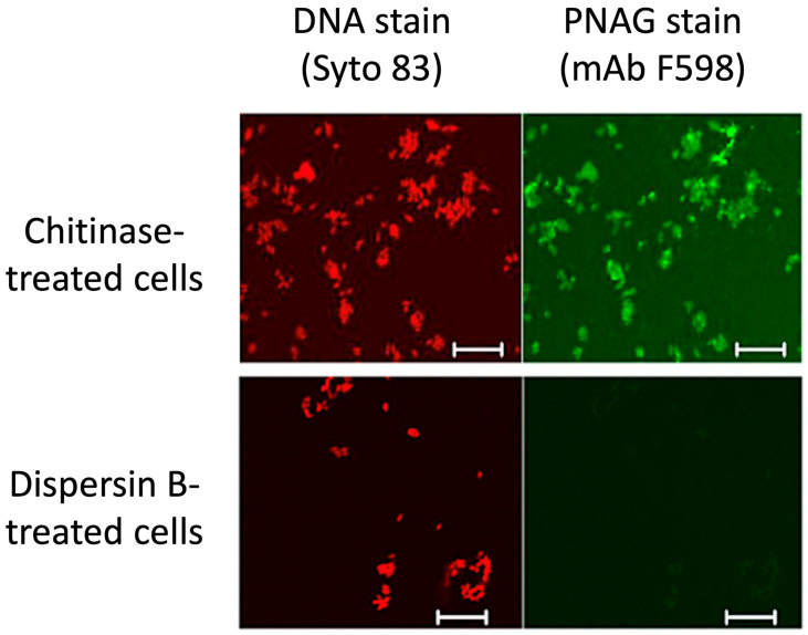
The extracellular matrix of most bacterial biofilms contains polysaccharides, proteins, and nucleic acids. These biopolymers have been shown to mediate fundamental biofilm-related phenotypes including surface attachment, intercellular adhesion, and biocide resistance. Enzymes that degrade polymeric biofilm matrix components, including glycoside hydrolases, proteases, and nucleases, are useful tools for studying the structure and function of biofilm matrix components and are also being investigated as potential antibiofilm agents for clinical use. Dispersin B is a well-studied, broad-spectrum antibiofilm glycoside hydrolase produced by Aggregatibacter actinomycetemcomitans. Dispersin B degrades poly-N-acetylglucosamine, a biofilm matrix polysaccharide that mediates biofilm formation, stress tolerance, and biocide resistance in numerous Gram-negative and Gram-positive pathogens. Dispersin B has been shown to inhibit biofilm and pellicle formation; detach preformed biofilms; disaggregate bacterial flocs; sensitize preformed biofilms to detachment by enzymes, detergents, and metal chelators; and sensitize preformed biofilms to killing by antiseptics, antibiotics, bacteriophages, macrophages, and predatory bacteria. This review summarizes the results of nearly 100 in vitro and in vivo studies that have been carried out on dispersin B since its discovery 20 years ago. These include investigations into the biological function of the enzyme, its structure and mechanism of action, and its in vitro and in vivo antibiofilm activities against numerous bacterial species. Also discussed are potential clinical applications of dispersin B.

Keywords: DspB; EPS; PIA; PNAG; Staphylococcus aureus; Staphylococcus epidermidis; biofilm matrix; biomaterial coating; eDNA; exopolysaccharide; extracellular DNA; matrix-degrading enzyme.


Conflict of interest statement

J.B.K. serves as an advisor for, owns equity in, and receives royalties from Kane Biotech Inc., Winnipeg, MB, Canada. This company is developing antibiofilm applications related to dispersin B. M.S. is an employee of Kane Biotech Inc., manufacturer of dispersin B (DispersinB®), and owns company stocks and stock options.

Figures

Figure 1
Figure 1
Dispersal of isolated Aggregatibacter actinomycetemcomitans biofilm colonies growing on the surface of polystyrene Petri dishes: (left panel) wild-type strain CU1000; (right panel) ΔdspB mutant strain JK1023. Satellite colonies surrounding the dispersed CU1000 biofilm colony were absent in the JK1023 culture. Photos were taken 3 d after inoculation. Scale bar = 1 mm. Image from [12].
Figure 2
Figure 2
Phylogenetic relatedness of dispersin B homologues based on pairwise alignments of the amino acid sequences listed in Table 1. The alignment was generated using ClustalW, and the phylogenetic tree was generated using FastTree software. Lacto-N-biosidase from Lactococcus lactis (GenBank accession number AGY45663.1) was used as an outgroup to locate the root of the tree. Horizontal branch lengths are proportional to the number of amino acid differences in the pairwise alignments. Bacterial families are indicated on the right.
Figure 3
Figure 3
Ribbon diagram of A. actinomycetemcomitans dispersin B; α-helices are colored red and green; β-strands are colored blue. Image source: Wikimedia Commons.
Figure 4
Figure 4
Dispersin B’s active site and mechanism of action: (A) Electrostatic surface potential at the active site showing the negatively charged amino acids (Asp56, Asp183, Glu184, Glu332), which create a shallow anionic region in the catalytic pocket. The size of the pocket is approximately 12 Å. GOL, glycerol; ACY, acetate. Figure generated using ChimeraX [29]. (B) Substrate hydrolysis mechanism proposed for dispersin B and other glycoside hydrolase family 20 hexosaminidases. In this substrate-assisted mechanism, Glu184 acts as the acid/base. The nucleophile is the N-acetyl group of the substrate, which is assisted by Asp183. Both exo- (dPNAG) and endoglycosidic (PNAG) cleavage are shown, where the leaving group is either deacetylated or acetylated, respectively. A suitably positioned Asp183 helps stabilize the oxazolium ion in the transition state. Figure generated using ChemDraw (PerkinElmer).
Figure 5
Figure 5
Confocal microscopic analysis of PNAG expression by Y. pestis strain KIM6+ grown at 28 °C overnight on Congo red agar. After treatment of bacterial cells with either chitinase (top panels) or dispersin B (bottom panels), cells were stained with Syto 83 to visualize DNA (red) and Alexa Fluor 488-conjugated mAb F598 to detect PNAG (green). Bars = 10 µm. Figure from Yoong et al. [40].
Figure 6
Figure 6
Abiotic surfaces coated with dispersin B resist S. epidermidis biofilm formation and surface attachment: (A) Biofilm formation by S. epidermidis strain NJ9709 on glass slides containing an ultrathin layered poly(allylamine hydrochloride) (PAH) hydrogel coating (left panel) or a PAH coating loaded with dispersin B (right panel). Bacteria were cultured inside plastic cloning cylinders (5 mm internal diameter) that were attached to the slide with high-vacuum grease. After 12 h, the biofilms were rinsed, the cloning cylinders were removed, and the slides were photographed. The rings correspond to the footprints of the cloning cylinders. The biofilm appeared as a white film on the unloaded PAH layer, which was absent on the dispersin-B-loaded PAH layer. (B) Attachment of S. epidermidis strain ATCC35984 to uncoated stainless steel disks, or to disks coated with polydopamine- or plasma-based coatings with or without grafted dispersin B. Source: (A) [65]; (B) redrawn from [66].
Figure 7
Figure 7
Effects of dispersin B on plant-associated bacteria: (A) Xanthomonas citri subsp. citri strain 306 forms aggregates when cultured in broth (left panel). These aggregates were rapidly dissolved upon dispersin B treatment (right panel). (B) Biofilm formation by Ralstonia solanacearum strain Molk2 in polystyrene microtiter plates in the absence or presence of 20 µg/mL dispersin B. Biofilms were stained with crystal violet. (C) Binding of Pseudomonas fluorescens strain WCS365 to tomato roots in the absence or presence of 20 µg/mL dispersin B. Bacteria were mixed with 6-day-old tomato roots for 90 min. The roots were then crushed, mixed by vortex agitation, diluted, and plated on agar for CFU enumeration. Each data point represents one individual root. (D) Tobacco leaves infected with Pectobacterium carotovorum subsp. carotovorum strain ATCC 15713. Leaves were photographed 24 h after inoculation: (left) wild-type tobacco leaf; (right) leaf from a transgenic tobacco plant expressing dispersin B. Source: (AC) J.B. Kaplan, unpublished data; (D) Ragunath et al. [104], N. Ramasubbu, unpublished data.

References

    1. Penesyan A., Paulsen I.T., Kjelleberg S., Gillings M.R. Three faces of biofilms: A microbial lifestyle, a nascent multicellular organism, and an incubator for diversity. NPJ Biofilms Microbiomes. 2021;7:80. doi: 10.1038/s41522-021-00251-2. - DOI - PMC - PubMed
    1. Penesyan A., Gillings M., Paulsen I.T. Antibiotic discovery: Combatting bacterial resistance in cells and in biofilm communities. Molecules. 2015;20:5286–5298. doi: 10.3390/molecules20045286. - DOI - PMC - PubMed
    1. Abdelhamid A.G., Yousef A.E. Combating bacterial biofilms: Current and emerging antibiofilm strategies for treating persistent infections. Antibiotics. 2023;12:1005. doi: 10.3390/antibiotics12061005. - DOI - PMC - PubMed
    1. Jiang Y., Geng M., Bai L. Targeting biofilms therapy: Current research strategies and development hurdles. Microorganisms. 2020;8:1222. doi: 10.3390/microorganisms8081222. - DOI - PMC - PubMed
    1. Kaplan J.B. Therapeutic potential of biofilm-dispersing enzymes. Int. J. Artif. Organs. 2009;32:545–554. doi: 10.1177/039139880903200903. - DOI - PubMed

MeSH terms

Substances

LinkOut - more resources