. 2024 Aug 1;16(8):1023.
doi: 10.3390/pharmaceutics16081023.

In Vitro Studies to Evaluate the Intestinal Permeation of an Ursodeoxycholic Acid-Conjugated Oligonucleotide for Duchenne Muscular Dystrophy Treatment

Affiliations

In Vitro Studies to Evaluate the Intestinal Permeation of an Ursodeoxycholic Acid-Conjugated Oligonucleotide for Duchenne Muscular Dystrophy Treatment

Marika Faiella et al. Pharmaceutics. .

Abstract

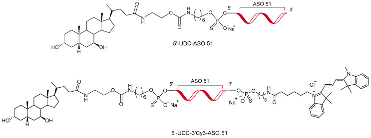
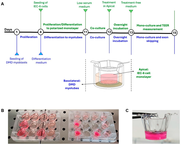
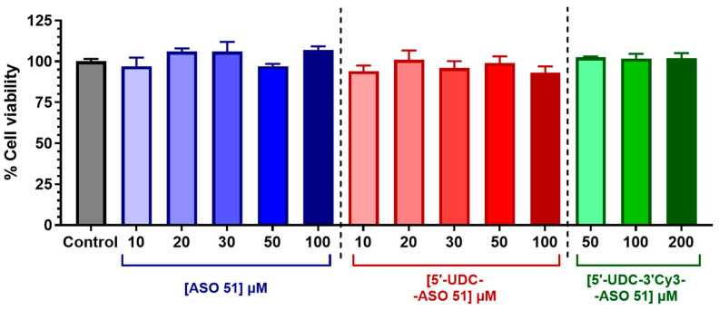
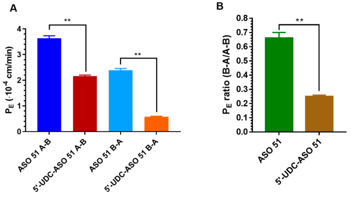
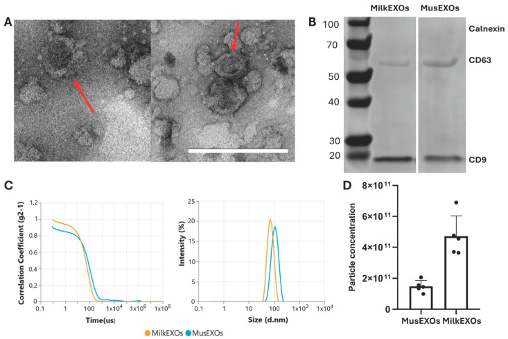
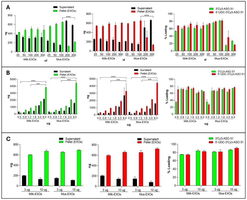
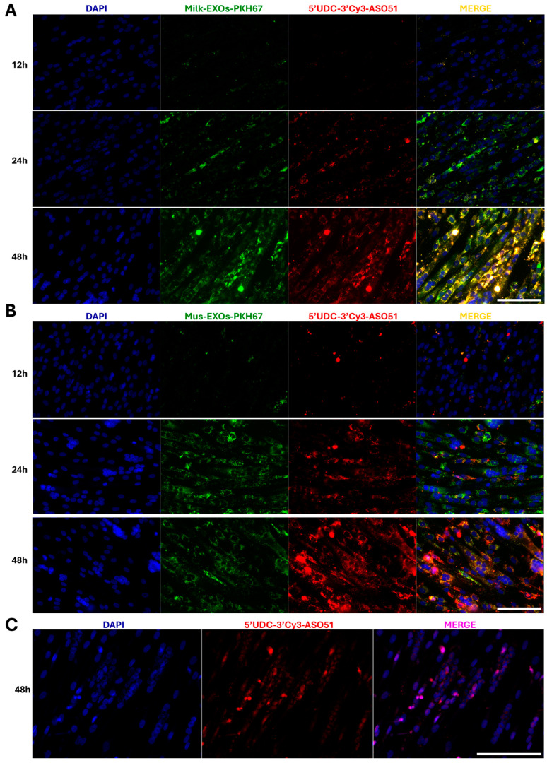
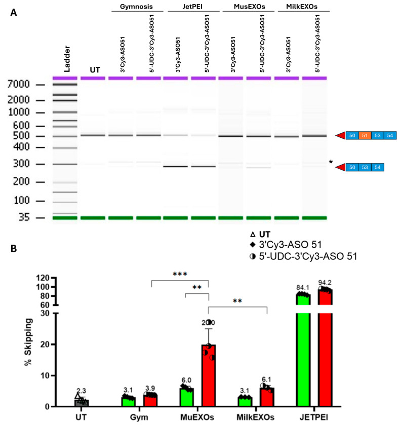
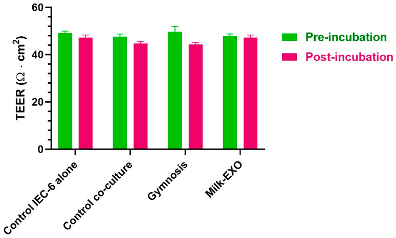
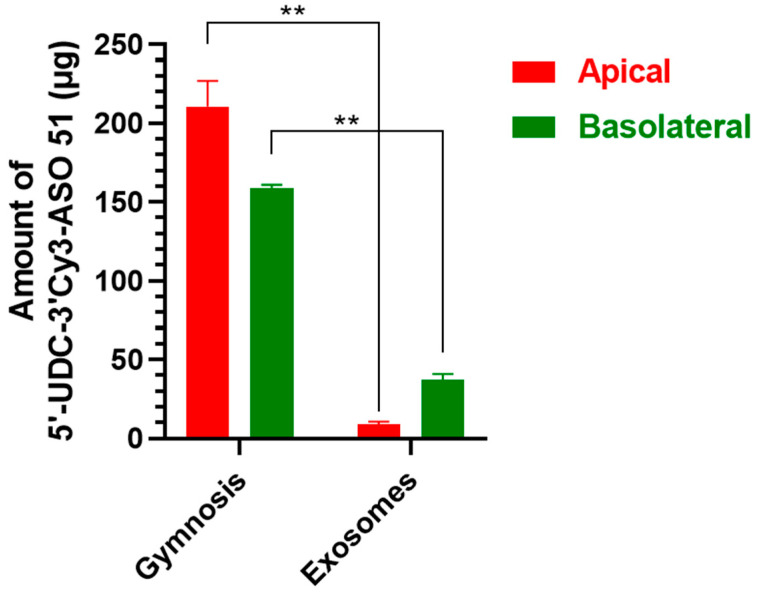
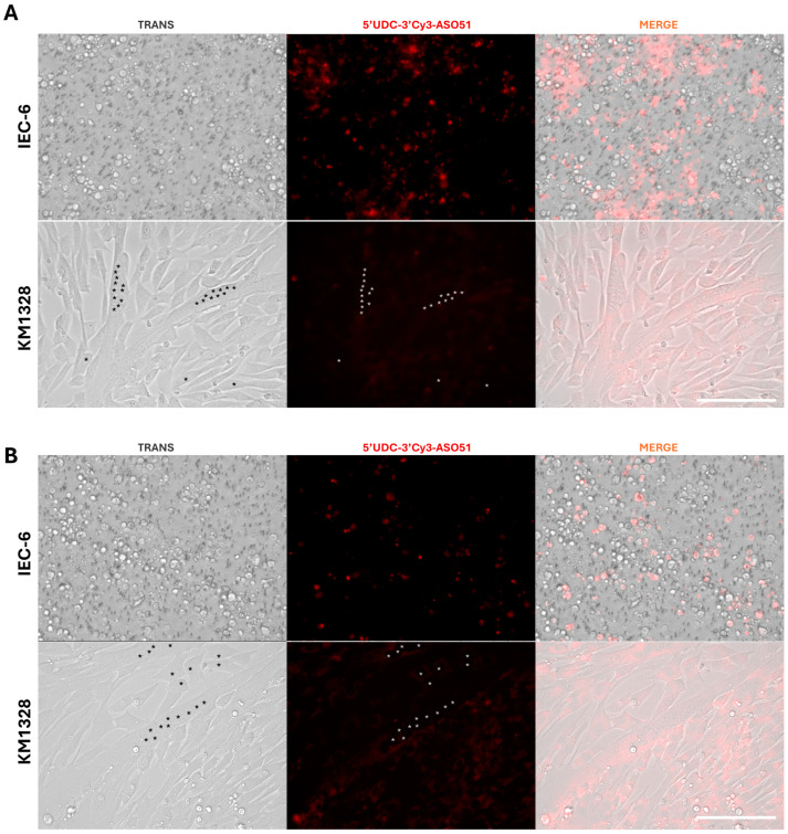
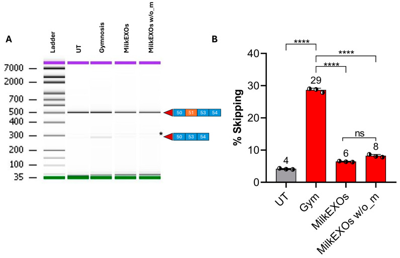
Delivery represents a major hurdle to the clinical advancement of oligonucleotide therapeutics for the treatment of disorders such as Duchenne muscular dystrophy (DMD). In this preliminary study, we explored the ability of 2'-O-methyl-phosphorothioate antisense oligonucleotides (ASOs) conjugated with lipophilic ursodeoxycholic acid (UDCA) to permeate across intestinal barriers in vitro by a co-culture system of non-contacting IEC-6 cells and DMD myotubes, either alone or encapsulated in exosomes. UDCA was used to enhance the lipophilicity and membrane permeability of ASOs, potentially improving oral bioavailability. Exosomes were employed due to their biocompatibility and ability to deliver therapeutic cargo across biological barriers. Exon skipping was evaluated in the DMD myotubes to reveal the targeting efficiency. Exosomes extracted from milk and wild-type myotubes loaded with 5'-UDC-3'Cy3-ASO and seeded directly on DMD myotubes appear able to fuse to myotubes and induce exon skipping, up to ~20%. Permeation studies using the co-culture system were performed with 5'-UDC-3'Cy3-ASO 51 alone or loaded in milk-derived exosomes. In this setting, only gymnotic delivery induced significant levels of exon skipping (almost 30%) implying a possible role of the intestinal cells in enhancing delivery of ASOs. These results warrant further investigations to elucidate the delivery of ASOs by gymnosis or exosomes.

Keywords: DMD myotubes; Duchenne muscular dystrophy; IEC-6 cells; antisense oligonucleotides; co-culture; conjugated oligonucleotides; exon skipping; exosomes; intestinal permeation; ursodeoxycholic acid.


Conflict of interest statement

The authors declare no conflicts of interest.

Figures

Figure 1
Figure 1
Chemical structure of 5′-UDC-ASO 51 [25] and 5′-UDC-3′Cy3-ASO 51.
Figure 2
Figure 2
Experimental design. (A) Schematic illustration of the step-by-step experimental design utilized in the non-contact co-culture experiment. Treatment = incubation with 100 µM (about 400 µg) 5′-UDC-3′Cy3-ASO 51 as free compound (gymnosis) or loaded in MilkEXOs. (B,C) Non-contact co-culture methodology used with cell culture inserts, highlighting the technical setup and arrangement of the cell culture system; the photos were acquired after the overnight incubation of 5′-UDC-ASO 51 as free compound or loaded in MilkEXO.
Figure 3
Figure 3
Cell viability of IEC-6 cells incubated overnight with different concentrations of ASO 51 (shades of blue), 5′-UDC-ASO 51 (shades of red) or 5′-UDC-3′Cy3-ASO 51 (shades of green). Results are reported as cell viability percentage (%) normalized to untreated control (in the absence of compounds). Data are expressed as mean ± S.E.M. of four independent experiments and were statistically analyzed by one-way ANOVA followed by Dunnett’s multiple comparisons test, showing no statistically significant differences in cell viability in the presence of compounds compared to untreated control.
Figure 4
Figure 4
(A) Permeation coefficients (PE) of ASO 51 and 5′-UDC-ASO 51 across the intestinal monolayer of IEC-6 cells from apical to basolateral compartments (AB) and vice versa (BA). All data are reported as the mean ± S.E.M of three independent experiments. (B) PE ratio between efflux (BA) and influx (AB) permeation of ASO 51 and 5′-UDC-ASO 51 across the intestinal monolayer of IEC-6 cells. The values are reported as the mean ± S.E.M. of three independent experiments. ** p < 0.0001.
Figure 5
Figure 5
Exosome characterization. (A) Morphological characterization of isolated exosomes was carried out using TEM, (left) MusEXOs, (right) MilkEXOs. Scale bar: 200 nm. Red arrows indicate exosomes. (B) Western blot analysis of exosome-specific markers (CD9 and CD63) and the endoplasmic reticulum marker (calnexin) was performed from exosomal protein extracts. (C) Size distribution of isolated exosomes measured using Zetasizer Ultra red instrument (Malvern Panalytical). The analysis produced a correlogram (left) and showed a distribution in the size of isolated exosomes with defined peaks and absence of polydispersity (right). (D) Particle concentration was evaluated by particle count, using the Zetasizer Ultra Red Instrument (Malvern Analytical). Each dot represents a different extraction of exosomes from the same initial amount of sample.
Figure 6
Figure 6
Co-incubation loading studies to identify the best ratio of ASOs and EXOs to achieve the highest loading efficiency. (A) Evaluation of the effect of different co-incubation volumes on loading efficiency. Different volumes (25, 50, 100 200 and 300 μL) of PBS were tested by mixing 3 μg of Milk-EXOs and Mus-EXOs with 800 ng of 5′-UDC-3′Cy3-ASO 51 (red) or 3′-Cy3-ASO 51 (green); (B) Evaluation of the effect of different amounts of ASOs on loading efficiency: 3 μg of Milk-EXOs and Mus-EXOs were mixed with 500, 800, 1200, 1600, 3000 and 6000 ng of 5′-UDC-3′Cy3-ASO 51 (red) or 3′Cy3-ASO 51 (green) for a ratio of 6:1, 3.75:1 2.5:1, 1.87:1, 1:1 and 1:2; (C) Evaluation of the effect of different quantity of EXOs on loading efficiency: 3 and 10 μg of Milk-EXOs and Mus-EXOs with 800 ng of 5′-UDC-3′Cy3-ASO 51 (red) or 3′Cy3-ASO 51 (green) in 100 μL of filtered 1X PBS. SUR-NATANT: ASOs quantified in the supernatant (BLACK); PELLET: ASOs quantified in the pellet, then loaded into the exosomes (GREEN: 3′Cy3-ASO 51; RED 5′-UDC-3′Cy3-ASO 51). Values are expressed as the mean ± standard deviation (SD) of three experiments. *** p < 0.0002 paired t-test, ordinary one-way ANOVA and two-way ANOVA multiple comparisons; **** p < 0.0001 paired t-test, ordinary one-way ANOVA and two-way ANOVA multiple comparisons.
Figure 7
Figure 7
EXOs-ASOs complexes fusion and distribution studies. Exosomes isolated from MilkEXOs (A) and MusEXOs (B) labelled with the fluorescent dye PKH67 (green) and loaded with 5′-UDC-3′Cy3-ASO 51 (red) were administered to KM1328 myotubes. The ability of EXOs–ASOs complexes to fuse with target cells and deliver ASOs was evaluated at different time points (12, 24 and 48 h). DAPI: Nucleus (blue). (C) 5′-UDC-3′Cy3-ASO 51 (red) was transfected with JetPEI into KM1328 myotubes. DAPI: Nucleus (blue). Scale bar: 75 μm.
Figure 8
Figure 8
Exon skipping analysis in KM1328 myotubes treated with Mus- or Milk-EXOs-ASOs complexes compared to gymnosis delivery and JetPEI transfection. (A) Representative gel capillary electrophoresis: on the right side of the image, the squares represent the exon composition of the corresponding bands of 514 bp for the out-of-frame transcript that includes exon 51 and 281 bp for the skipped transcript. Purple: Upper marker; green: Lower marker. * Unspecific band highlighted in each sample, including UT. (B) Exon skipping results: the quantification of exon skipping was performed by determining the percentage ratio, calculated as the area of the skipped transcript divided by the total area of both skipped and unskipped transcripts, multiplied by 100. UT: Untreated; Gym: Gymnosis. Values are expressed as the mean ± standard deviation (SD) of four experiments. ** p < 0.001 paired t-test and ordinary one-way ANOVA; *** p < 0.0002 paired t-test and ordinary one-way ANOVA.
Figure 9
Figure 9
TEER measurement of IEC-6 cell monolayer co-cultured for 24 h with myotubes measured before (“pre-incubation”) and after (“post-incubation”) the incubation with 5′-UDC-3′Cy3-ASO 51 in gymnotic delivery (“Gymnosis”) or as MilkEXO (“MilkEXO”). IEC-6 cell monolayer alone or co-cultured 24 h with myotubes were used as negative controls. Data are expressed as the mean ± S.E.M. of three independent experiments. No significant differences were observed.
Figure 10
Figure 10
Amounts of 5′-UDC-3′Cy3-ASO 51 expressed in µg contained in 0.4 mL of medium for the apical compartment and 2 mL of medium for the basolateral compartment, quantified 48 h after the end of the co-culture experiment. Data are expressed as the mean ± S.E.M. of three independent experiments. ** p < 0.01 in the comparison of the same compartment in different incubation conditions.
Figure 11
Figure 11
Fluorescence distribution of 5′-UDC-3′Cy3-ASO 51 into the IEC-6 and KM1328 myotubes after 48 h of gymnotic delivery (A) or incapsulated into MilkEXOs (B). With *, we highlight the nuclei of the myotubes. Scale bar: 75 μm.
Figure 12
Figure 12
Exon skipping analysis in in vitro permeation studies. (A) Representative gel capillary electrophoresis: on the right side of the image, the squares represent the exon composition of the corresponding bands as described in Figure 8. Purple: Upper marker; green: Lower marker. * Unspecific band highlighted in each sample, including UT. (B) Exon skipping quantification. UT: Untreated; Gym: Gymnosis. MilkEXOs w/o_m: exon skipping results from the direct administration of MilkEXOs-5′-UDC-3′Cy3-ASO 51 to the myotubes. Values are expressed as the mean ± standard deviation (SD) of three experiments. **** p < 0.0001 (paired t-test and one-way ANOVA).

References

    1. Duan D., Goemans N., Takeda S., Mercuri E., Aartsma-Rus A. Duchenne Muscular Dystrophy. Nat. Rev. Dis. Primers. 2021;7:13. doi: 10.1038/s41572-021-00248-3. - DOI - PMC - PubMed
    1. Fox H., Millington L., Mahabeer I., van Ruiten H. Duchenne Muscular Dystrophy. BMJ. 2020;368:l7012. doi: 10.1136/bmj.l7012. - DOI - PubMed
    1. Vinjamuri B.P., Pan J., Peng P. A Review on Commercial Oligonucleotide Drug Products. J. Pharm. Sci. 2024;113:1749–1768. doi: 10.1016/j.xphs.2024.04.021. - DOI - PubMed
    1. Bylo M., Farewell R., Coppenrath V.A., Yogaratnam D. A Review of Deflazacort for Patients with Duchenne Muscular Dystrophy. Ann. Pharmacother. 2020;54:788–794. doi: 10.1177/1060028019900500. - DOI - PubMed
    1. FDA Approves Nonsteroidal Treatment for Duchenne Muscular Dystrophy. [(accessed on 27 June 2024)]; Available online: https://www.fda.gov/news-events/press-announcements/fda-approves-nonster....

LinkOut - more resources