. 2024 Jul 26;16(8):1206.
doi: 10.3390/v16081206.

Characterization of Human Immortalized Keratinocyte Cells Infected by Monkeypox Virus

Affiliations

Characterization of Human Immortalized Keratinocyte Cells Infected by Monkeypox Virus

Chaode Gu et al. Viruses. .

Abstract

Monkeypox virus (MPXV) can induce systemic skin lesions after infection. This research focused on studying MPXV proliferation and the response of keratinocytes. Using transmission electron microscopy (TEM), we visualized different stages of MPXV development in human immortalized keratinocytes (HaCaT). We identified exocytosis of enveloped viruses as the exit mechanism for MPXV in HaCaT cells. Infected keratinocytes showed submicroscopic changes, such as the formation of vesicle-like structures through the recombination of rough endoplasmic reticulum membranes and alterations in mitochondrial morphology. Transcriptome analysis revealed the suppressed genes related to interferon pathway activation and the reduced expression of antimicrobial peptides and chemokines, which may facilitate viral immune evasion. In addition, pathway enrichment analysis highlighted systemic lupus erythematosus pathway activation and the inhibition of the Toll-like receptor signaling and retinol metabolism pathways, providing insights into the mechanisms underlying MPXV-induced skin lesions. This study advances our understanding of MPXV's interaction with keratinocytes and the complex mechanisms leading to skin lesions.

Keywords: HaCaT; RNA-seq; TEM; characterization; monkeypox virus.


Conflict of interest statement

The authors declare no conflicts of interest.

Figures

Figure 1
Figure 1
The phylogenetic analysis of hMpxV/China/GZ8H-01/2023. Phylogenetic analysis showed the hMpxV/China/GZ8H-01/2023 isolate belonged to clade IIb of MPXV, highlighted in red.
Figure 2
Figure 2
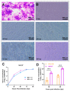
The growth dynamics of MPXV in HaCaT cells. (A) Cytopathic effects (CPE) of HaCaT cells infected with MPXV by crystal violet staining. (B) CPE of HaCaT cells infected with 0.5 MOI MPXV at different time points (0, 24, 48, 72, and 96 hpi). (C) HaCaT cells infected with 0.1 or 0.5 MOI MPXV, virus concentration in the supernatant were detected and recorded at 0, 24, 48, 72, and 96 hpi, respectively (n = 3). (D) Infection of Vero-E6 and HaCaT cells with 0.5 MOI MPXV, followed by virus concentration in the supernatant detection and recording at 24 and 48 hpi, respectively (n = 3). Data were analyzed by one-way ANOVA and t-test. p  <  0.05 was considered statistically significant. “***” represents p  <  0.001.
Figure 3
Figure 3
Morphogenesis of MPXV in HaCaT cells. (A) Numerous MPXV virions were observed in cytoplasmic matrix of the infected HaCaT cells. (B,C) The mature MPXV virions presented two different forms, the unencapsulated virion IMV and the encapsulated virion IEV. (D) The viral factory (marked with a yellow pentagram) encompassed various structures, including crescent structure (green triangle), immature (blue triangle) and mature (red triangle) viral particles, as well as multi-membrane vesicles (green asterisk), and abnormal mitochondria (blue asterisk). (E) Transformation of MPXV from viral crescent structures to IMV. (F) The mature virions were enveloped by membranes. (G) IEV at the periphery of the cell. (H) IEV was transported out of the cell through exocytosis. (I) The extracellular virions enveloped by singular membrane near the plasma membrane were termed as CEV. (J) Released extracellular virions were termed as EEV.
Figure 4
Figure 4
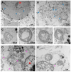
Ultrastructural features of HaCaT cells infected with MPXV. (A) The HaCaT cells at the early stage of MPXV infection exhibited a substantial presence of EEV attached to the cell membrane surface, along with a few virions within the envelope, while maintaining an intact mitochondrial structure. (B) Mitochondria in late-stage infected cells showed an enlarged and rounded morphology, accompanied by a reduction in or the disappearance of cristae, with the matrices filled with granular contents. (C) Different forms of spherical mitochondria showed varying degrees of damage to their inner and outer membranes or cristae. (D) Numerous vesicle structures derived from rER were observed in the infected HaCaT cells. (E) The moment when the rER membrane protruded outward to form a vesicle. (F) At the late stage of MPXV infection, abundant multi-membrane vesicle structures appeared in HaCaT cells, along with the presence of mature virions nearby. (G) The nucleus in a portion of infected HaCaT cells exhibited a C-type morphology, with an opening facing the region of MPXV aggregation.
Figure 4
Figure 4
Ultrastructural features of HaCaT cells infected with MPXV. (A) The HaCaT cells at the early stage of MPXV infection exhibited a substantial presence of EEV attached to the cell membrane surface, along with a few virions within the envelope, while maintaining an intact mitochondrial structure. (B) Mitochondria in late-stage infected cells showed an enlarged and rounded morphology, accompanied by a reduction in or the disappearance of cristae, with the matrices filled with granular contents. (C) Different forms of spherical mitochondria showed varying degrees of damage to their inner and outer membranes or cristae. (D) Numerous vesicle structures derived from rER were observed in the infected HaCaT cells. (E) The moment when the rER membrane protruded outward to form a vesicle. (F) At the late stage of MPXV infection, abundant multi-membrane vesicle structures appeared in HaCaT cells, along with the presence of mature virions nearby. (G) The nucleus in a portion of infected HaCaT cells exhibited a C-type morphology, with an opening facing the region of MPXV aggregation.
Figure 5
Figure 5
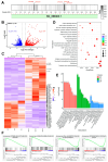
RNA-seq analyses of the MPXV-infected HaCaT cells. (A) The location of MPXV transcript in viral genome. (B) The volcano plot of DEGs regarding uninfected and 24 h post-infected HaCaT cells (Log2 (fold change) > 1, adjusted p < 0.05). The red dots represent upregulated genes while the blue dots represent downregulated genes. (C) The heat map of DEGs, in which the genes associated with interferon and IL17 signaling pathways were marked. (D) KEGG analysis of the DEGs. (E) DEGs were categorized based on their gene ontology (GO) terms related to biological process (BP), cell component (CC), and molecular function (MF). (F) Gene set enrichment analysis of specified gene sets, including lupus erythematosus signaling pathway, body assembly, Toll-like receptor signaling pathway, and retinoid metabolism.

References

    1. Foster S.O., Brink E.W., Hutchins D.L., Pifer J.M., Lourie B., Moser C.R., Cummings E.C., Kuteyi O.E., Eke R.E., Titus J.B., et al. Human monkeypox. Bull. World Health Organ. 1972;46:569–576. - PMC - PubMed
    1. Del Rio C., Malani P.N. Update on the Monkeypox Outbreak. JAMA. 2022;328:921–922. doi: 10.1001/jama.2022.14857. - DOI - PubMed
    1. Mitjà O., Ogoina D., Titanji B.K., Galvan C., Muyembe J.J., Marks M., Orkin C.M. Monkeypox. Lancet. 2023;401:60–74. doi: 10.1016/S0140-6736(22)02075-X. - DOI - PMC - PubMed
    1. Dou X., Li F., Ren Z., Zhang D., Li J., Li D., Sun Y., Jin H., Li R., Li W., et al. Clinical, epidemiological, and virological features of Mpox in Beijing, China—May 31–June 21, 2023. Emerg. Microbes Infect. 2023;12:2254407. doi: 10.1080/22221751.2023.2254407. - DOI - PMC - PubMed
    1. Rosa R.B., Ferreira de Castro E., Vieira da Silva M., Paiva Ferreira D.C., Jardim A.C.G., Santos I.A., Marinho M.d.S., Ferreira França F.B., Pena L.J. In vitro and in vivo models for monkeypox. iScience. 2023;26:105702. doi: 10.1016/j.isci.2022.105702. - DOI - PMC - PubMed

Publication types

Associated data

LinkOut - more resources