. 2024 Jul 29;16(8):1214.
doi: 10.3390/v16081214.

Interaction between SARS-CoV PBM and Cellular PDZ Domains Leading to Virus Virulence

Affiliations

Interaction between SARS-CoV PBM and Cellular PDZ Domains Leading to Virus Virulence

Jose M Honrubia et al. Viruses. .

Abstract

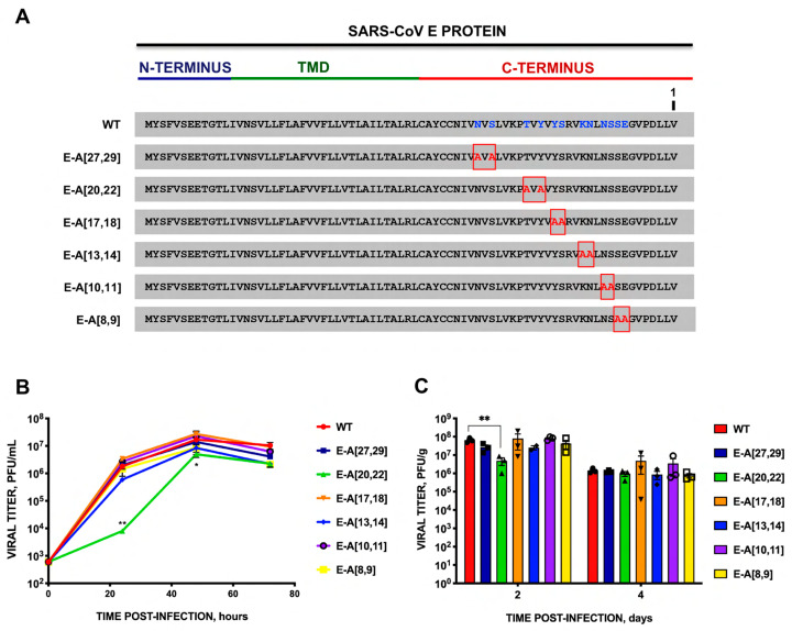
The interaction between SARS-CoV PDZ-binding motifs (PBMs) and cellular PDZs is responsible for virus virulence. The PBM sequence present in the 3a and envelope (E) proteins of SARS-CoV can potentially bind to over 400 cellular proteins containing PDZ domains. The role of SARS-CoV 3a and E proteins was studied. SARS-CoVs, in which 3a-PBM and E-PMB have been deleted (3a-PBM-/E-PBM-), reduced their titer around one logarithmic unit but still were viable. In addition, the absence of the E-PBM and the replacement of 3a-PBM with that of E did not allow the rescue of SARS-CoV. E protein PBM was necessary for virulence, activating p38-MAPK through the interaction with Syntenin-1 PDZ domain. However, the presence or absence of the homologous motif in the 3a protein, which does not bind to Syntenin-1, did not affect virus pathogenicity. Mutagenesis analysis and in silico modeling were performed to study the extension of the PBM of the SARS-CoV E protein. Alanine and glycine scanning was performed revealing a pair of amino acids necessary for optimum virus replication. The binding of E protein with the PDZ2 domain of the Syntenin-1 homodimer induced conformational changes in both PDZ domains 1 and 2 of the dimer.

Keywords: PBM-PDZ; coronavirus; virulence; virus–host interaction.


Conflict of interest statement

The authors declare no conflicts of interest. The funders had no role in the design of the study; in the collection, analyses, or interpretation of data; in the writing of the manuscript; or in the decision to publish the results.

Figures

Figure 1
Figure 1
Virulence of SARS-CoV mutants in which the 3a and E protein PBMs have been deleted or exchanged. (A) Scheme of mutants generated by combining mutations in the PBM of 3a (SVPL) and E (DLLV) proteins. Optimum viral titers are shown on the right. Mean weight loss (B) and survival (C) of 16-week-old infected Balb/c mice were monitored for 10 days and compared with those of mock-infected mice. Error bars represent the standard deviation of mouse weight for each experimental variable. Data shown are the mean and standard deviation obtained in three experiments. Uninfected mice, gray circles. Mice inoculated with 100,000 pfu of parental virus (WT, red), or with mutant viruses generated by reverse genetics, without the PBM of 3a protein (3a-PBM-, blue), without the PBM of E protein (E-PBM-, pink), without both motifs (3a-PBM-/E-PBM-, orange), with the PBM of 3a protein in the context of E protein (E-PBM3a, green) or with the PBM of both proteins swapped (3a-PBME/E-PBM3a, purple).
Figure 2
Figure 2
Potential structure of the active domain included within E protein that may be required for the PBM function. Two different structures were considered. One in which, in addition to the PBM core formed by the four carboxyterminal amino acids, E protein contained a hypothetic active domain located to the 5′ side of the PBM (top bar). A second one in which the two domains (PBM and active domain modulating the activity of the first one), overlapped at the carboxy-terminal end (bottom bar).
Figure 3
Figure 3
Growth kinetics of SARS-CoV mutants with double mutations of the polar residues of the carboxyterminal end of E protein replaced by alanine. (A) Sequence of the parental virus (WT) E protein, and that of the mutant viruses in which two polar residues of the carboxy-terminal end have been mutated to alanine (E-A[X,X], where X is the position of the mutated residue starting from the carboxyl-terminal end of E protein, indicated as 1 in the figure. (B) Vero E6 cells were infected at a moi of 0.001 with parental SARS-CoV (WT) (red circles) and with rSARS-CoV-E-A[27,29] (dark blue squares), E-A[20,22] (green triangles), E-A[17,18] (orange inverted triangles), E-A[13,14] (blue diamonds), E-A[10,11] (purple circles) or E-A[8,9] (yellow squares). Supernatants were collected at 24, 48, and 72 h post-infection (hpi) and titrated by the lysis plaque formation method. These results are derived from three independent experiments. (C) Groups of six 16-week-old Balb/c mice were intranasally inoculated with 100,000 pfu/mouse of the indicated rSARS-CoV. At 2 and 4 days post-infection (dpi), three mice per group were sacrificed, and viral titers in the lungs were analyzed. Error bars represent the standard error of the mean. * p-value < 0.05; ** p value < 0.01).
Figure 4
Figure 4
Virulence of SARS-CoV mutants with double substitutions of polar residues of the carboxyterminal E protein by alanine. Groups of five 16-week-old Balb/c mice were intranasally inoculated with DMEM (Mock, gray), or infected with 100.000 pfu/mouse of the parental virus (WT, red), or with the engineered rSARS-CoV: E-A[27,29] (dark blue), E-A[20,22] (green), E-A[17,18] (orange), E-A[13,14] (blue), E-A[10,11] (purple) or E-A[8,9] (yellow). (A) Weight loss and (B) survival were determined for 10 dpi. Vertical bars represent the standard error of the mean weight of the mice.
Figure 5
Figure 5
Growth kinetics of SARS-CoV mutants with amino acid substitutions by glycine in carboxyterminal E protein residues that may be relevant in the interaction of the E PBM with cellular Syntenin-1, according to the developed in silico model. (A) The E protein sequence of the parental virus (WT) and that of the glycine substitution mutants engineered is shown. (E-G[X], where X is the position of the mutated residue from the carboxy-terminal end of E protein, numbered as in Figure 3A. (B) Vero E6 cells were infected at a moi of 0.001 with parental SARS-CoV (WT) (red circles) and with mutants E-G[1] (dark blue square), E-G[2] (green triangle), E-G[3] (orange inverted triangle), E-G[4] (blue diamond), E-A[5,6] (yellow square), E-G[8,9,10] (brown inverted triangle) or with E-G[1,2,3,4] (pink circle). Culture supernatants were collected at 24, 48, and 72 hpi, and titrated by plaque assays. These results are derived from three independent experiments. (C) Groups of six 16-week-old Balb/c mice were intranasally inoculated with 100,000 pfu/mouse of rSARS-CoVs. At 2 and 4 dpi, three mice per group were sacrificed, and viral titer in the lung was analyzed. Vertical bars represent the standard error of the mean. **, p-value < 0.01.
Figure 6
Figure 6
Virulence of SARS-CoV mutants with glycine substitutions in the carboxy-terminal residues of the E protein that may participate in the interaction of this protein with Syntenin-1 according to the structure proposed by the in silico model. Groups of five 16-week-old Balb/c mice were intranasally inoculated with DMEM (Mock, gray), or with 100,000 pfu/mouse of the parental SARS-CoV virus (WT), or with the glycine replacement mutants generated: E-G[1] (dark blue), E-G[2] (green), E-G[3] (orange), E-G[4] (blue), E-G[5,6] (yellow), E-G[8,9,10] (brown), E-G[1,2,3,4] (pink). Weight loss (A) and survival (B) were monitored for 10 dpi. Vertical bars represent the standard error of the mean weight of the mice.
Figure 7
Figure 7
Effect of carboxyl-terminal binding of SARS-CoV E protein on the structure of Syntenin-1. The left panel shows free Syntenin-1 (blue) with regions that change colored by their variance (orange < orange-red < red) calculated by THESEUS, with both PDZ2 domains indicated by arrows. The right panel shows free Syntenin-1 (blue) superposed to the bound form (yellow).
Figure 8
Figure 8
Proposed conformation of the binding between the carboxy-terminus of E protein and Syntenin-1. At the top, the PDZ2 domain of human Syntenin-1 is depicted in yellow, with the amino acids involved in binding to E protein in brown. The carboxyl terminus of SARS-CoV E protein is shown in blue and the four main residues of the PBM are circled in red. Hydrogen bridges are depicted in red and the contacts between the residues of both proteins are in cyan. The same conformation is shown schematically at the bottom. The last 10 residues of the carboxyl-terminal protein are located in the center of the pocket of the PDZ2 domain of Syntenin-1. Direct contacts between the residues are shown in blue and hydrogen bonds in black.
Figure 9
Figure 9
Predicted functional motifs in SARS-CoV E protein affected by alanine substitution in polar residues. The full-length sequence and structural domains of SARS-CoV E protein are shown in the top bar. In the left column, different predicted functional motifs in the E protein of SARS-CoV are indicated [35]. Color coding of amino acids (aa): in red, small hydrophobic aa (including aromatic aa except for tyrosine); in blue: acidic aa; in magenta: basic aa (except histidine); in green, aa with hydroxyl, thiol, or amine groups (glycine is also included). The location of the functional motifs identified in the E protein and the mutants with two polar residues at the carboxyl end substituted by alanine (E-A[X,X], where X is the position of the mutated residue starting at the carboxyl-terminal end of the E protein, are shown. Vertical colored rectangles indicate the location of predicted SARS-CoV-E protein motifs.

References

    1. Gorbalenya A.E., Baker S.C., Baric R.S., de Groot R.J., Drosten C., Gulyaeva A.A., Haagmans B.I., Lauber C., Leontovich A.M., Neuman B.W., et al. The species Severe acute respiratory syndrome-related coronavirus: Classifying 2019-nCoV and naming it SARS-CoV-2. Nat. Microbiol. 2020;5:536–544. - PMC - PubMed
    1. Drosten C., Gunther S., Preiser W., van der Werf S., Brodt H.R., Becker S., Rabenau H., Panning M., Kolesnikova L., Fouchier R.A., et al. Identification of a novel coronavirus in patients with severe acute respiratory syndrome. N. Engl. J. Med. 2003;348:1967–1976. doi: 10.1056/NEJMoa030747. - DOI - PubMed
    1. Rota P.A., Oberste M.S., Monroe S.S., Nix W.A., Campganoli R., Icenogle J.P., Peñaranda S., Bankamp B., Maher K., Chen M.-H., et al. Characterization of a novel coronavirus associated with severe acute respiratory syndrome. Science. 2003;300:1394–1399. doi: 10.1126/science.1085952. - DOI - PubMed
    1. Kennedy M.B. Origin of PDZ (DHR, GLGF) domains. Trends Biochem. Sci. 1995;20:350. doi: 10.1016/S0968-0004(00)89074-X. - DOI - PubMed
    1. Castaño-Rodriguez C., Honrubia J.M., Gutiérrez-Álvarez J., Sola I., Enjuanes L. Viral PDZ Binding Motifs Influence Cell Behavior Through the Interaction with Cellular Proteins Containing PDZ Domains. Methods Mol. Biol. 2021;2256:217–236. - PubMed

Publication types

MeSH terms

Substances

Associated data

LinkOut - more resources