. 2024 Aug 1;16(8):1239.
doi: 10.3390/v16081239.

An ISG15-Based High-Throughput Screening Assay for Identification and Characterization of SARS-CoV-2 Inhibitors Targeting Papain-like Protease

Affiliations

An ISG15-Based High-Throughput Screening Assay for Identification and Characterization of SARS-CoV-2 Inhibitors Targeting Papain-like Protease

Subodh Kumar Samrat et al. Viruses. .

Abstract

Emergence of newer variants of SARS-CoV-2 underscores the need for effective antivirals to complement the vaccination program in managing COVID-19. The multi-functional papain-like protease (PLpro) of SARS-CoV-2 is an essential viral protein that not only regulates the viral replication but also modulates the host immune system, making it a promising therapeutic target. To this end, we developed an in vitro interferon stimulating gene 15 (ISG15)-based Förster resonance energy transfer (FRET) assay and screened the National Cancer Institute (NCI) Diversity Set VI compound library, which comprises 1584 small molecules. Subsequently, we assessed the PLpro enzymatic activity in the presence of screened molecules. We identified three potential PLpro inhibitors, namely, NSC338106, 651084, and 679525, with IC50 values in the range from 3.3 to 6.0 µM. These molecules demonstrated in vitro inhibition of the enzyme activity and exhibited antiviral activity against SARS-CoV-2, with EC50 values ranging from 0.4 to 4.6 µM. The molecular docking of all three small molecules to PLpro suggested their specificity towards the enzyme's active site. Overall, our study contributes promising prospects for further developing potential antivirals to combat SARS-CoV-2 infection.

Keywords: FRET assay; ISG15; PLpro; SARS-CoV-2; high-throughput screening.


Conflict of interest statement

A patent has been filed for this study. The authors declare no conflicts of interest.

Figures
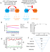
Figure 1
Figure 1
Measurements of PLpro activity and HTS using the ISG15-based assay. (A) The ISG15 FRET assay relies on FRET from CFP to YFP. In our construct, CFP and YFP, the FRET pairs, are fused together with the ISG15 protein, which is sensitive to PLpro cleavage. Consequently, the natural FRET signal can be disrupted when the PLpro enzyme cleaves and separates CFP-ISG15 from YFP. (B) The ratio of fluorescence at 530/475 is shown for different conditions: PLpro alone (blue), CFP-3C-ISG15-YFP (CIY) substrate alone (red), CIY substrate with PLpro (green), and CIY substrate with PLpro and GRL0617 (purple). (C) The kinetic analysis of PLpro and the CIY substrate. n = 3. (D) The Z’ score, S/B ratio, and CV for HTS. (E) The screening flowchart.
Figure 2
Figure 2
Identified candidate PLpro inhibitors. (A) Structures of GRL0617 and the identified small molecule inhibitors: NSC207895, 338106, 341956, 651084, and 679525. (B) Dose–response curves for small molecule inhibitors tested against PLpro using the ISG15-based assay. The ISG15−based CIY substrate was employed, and its cleavage by PLpro was assessed in the presence of small molecule inhibitors in the presence or absence of 0.01% Tween−20. GRL0617 served as the positive control inhibitor, and the IC50 of each inhibitor was determined. All experiments were performed in triplicate, and all data are expressed as the mean ± standard deviation.
Figure 3
Figure 3
Antiviral activities. (A) Curve fitting of data from the plaque reduction assay of small molecule PLpro inhibitors against SARS-CoV-2. Dose–response titrations were carried out in 96-well plates with Vero-E6 cells (1.5 × 10⁴ cells/well). On the following day, the cells were infected with SARS-CoV-2 (MOI: 0.01) while exposed to increasing concentrations of the compounds (ranging from 25 μM to 0.3 μM). Virus infection was quantified using a plaque reduction assay after 24 h. The signals from the samples were normalized using signals from the DMSO control wells. The data represent the mean ± standard error from triplicate experiments, N = 3. (B) Representative images of immunofluorescence assay (IFA) for dose-dependent inhibition of the Washington Strain (WA1) of SARS-CoV-2 treated with decreasing concentrations (50 µM to 3.1 µM) of NSC338106, 651084, and 679525. Vero−E6 cells were infected with the virus, treated with compounds at indicated concentrations for 24 h, fixed, and immunolabeled with a primary SARS-CoV-2 nucleocapsid monoclonal antibody and a goat anti−mouse secondary Alexa−488 antibody. (C) Dose−dependent inhibition of protein expression of the WA1 of SARS-CoV-2 treated with decreasing concentrations (50 µM to 3.1 µM) of compounds. Normalized data obtained from IFA as shown in panel (B) was plotted against concentration of compounds. The intensities of Alexa−488 positive cells for the DMSO control were set as 100%, N = 6.
Figure 4
Figure 4
Inhibition of the Omicron strain of SARS-CoV-2. (A) Immunofluorescence assay (IFA) for dose-dependent inhibition of the Omicron strain of SARS-CoV-2 treated with decreasing concentrations (50 µM to 1.6 µM) of compounds. Vero-E6 cells were infected with the virus, treated with compounds at indicated concentrations for 48 h, fixed, and immunolabeled with a primary SARS-CoV-2 nucleocapsid monoclonal antibody and a goat anti-mouse secondary Alexa-488 antibody. (B) Quantification of the IFA data shown in panel (A). Normalized data obtained from IFA was plotted against concentration of compounds. The intensities of Alexa-488-positive cells for the DMSO control were set as 100%, N = 3.
Figure 5
Figure 5
Cytotoxicity activity of NSC338106, 651084, and 679525. Vero−E6 cells were incubated with various concentrations of compounds, and then viability was assayed at 48 h of incubation, using the WST assay, N = 3.
Figure 6
Figure 6
Inhibition of the CIY substrate cleavage by the SARS-CoV-2 PLpro inhibitors. A total of 1 µM of the PLpro protein was incubated for 30 min with increasing concentrations of each inhibitor separately in a Tris buffer. After a 30 min incubation, the CIY protein substrate was added and further incubated for one hour, followed by SDS−PAGE analysis. GRL0617 was used as the positive control. The uncleaved substrate corresponds to 70 kDa, and the cleaved product is 47 kDa. The remaining bands are impurities of protein. All inhibitors were used at 60 µM, 30 µM, and 15 µM concentrations. Gel quantification is shown in the lower panel, N = 4.
Figure 7
Figure 7
Surface plasmon resonance (SPR) for binding affinity of compounds to recombinant SARS-CoV-2 PLpro. Equilibrium dissociation constant (KD) values were determined using both the 1:1 Langmuir kinetic model and the steady−state affinity model, which measure the respective association (Ka) and dissociation (Kd) rates of each compound to fit the data from titrating 100−0.1 μM for NSC338106 (A), NSC651084 (B), and NSC679525 (C) against immobilized PLpro.
Figure 8
Figure 8
Mechanism for inhibition of SARS-CoV-2 PLpro. Lineweaver–Burk plots for inhibition of the SARS-CoV-2 PLpro by NSC651084, NSC338106, and NSC679525. The SARS-CoV-2 PLpro at 40 nM was mixed with DMSO, NSC651084, NSC338106, and NSC679525 with varying concentrations. Peptide-based AMC substrate was added at various concentrations (1 μM−60 μM).
Figure 9
Figure 9
Binding models of candidate inhibitors to the SARS-CoV-2 PLpro in the putative ligand binding sites. (A) Overall locations of the three binding sites are as follows: the active site is colored yellow, allosteric site 1 is colored green, and allosteric site 2 is colored magenta. (B) Detailed molecular interactions between inhibitors and predicted binding site of PLpro. NSC338106 is docked into allosteric site 1, NSC651084 is docked into the active site, and NSC679525 is docked into allosteric site 2. The right panels show 2D interaction maps where π–π interactions are indicated in green, π–cation interactions in red, salt bridges in gradient blue-red, and hydrogen bonding in purple, with arrows indicating the role as donor or acceptor. The left panels display 3D interactions, with dashed lines indicating different interaction types by color: yellow for hydrogen bonding, purple for salt bridges, green for π–cation interactions, and cyan for π–π interactions.

References

    1. Lipsitch M., Krammer F., Regev-Yochay G., Lustig Y., Balicer R.D. SARS-CoV-2 breakthrough infections in vaccinated individuals: Measurement, causes and impact. Nat. Rev. Immunol. 2022;22:57–65. doi: 10.1038/s41577-021-00662-4. - DOI - PMC - PubMed
    1. Puhach O., Adea K., Hulo N., Sattonnet P., Genecand C., Iten A., Jacquérioz F., Kaiser L., Vetter P., Eckerle I., et al. Infectious viral load in unvaccinated and vaccinated individuals infected with ancestral, Delta or Omicron SARS-CoV-2. Nat. Med. 2022;28:1491–1500. doi: 10.1038/s41591-022-01816-0. - DOI - PubMed
    1. Flacco M.E., Acuti Martellucci C., Baccolini V., De Vito C., Renzi E., Villari P., Manzoli L. COVID-19 vaccines reduce the risk of SARS-CoV-2 reinfection and hospitalization: Meta-analysis. Front. Med. 2022;9:1023507. doi: 10.3389/fmed.2022.1023507. - DOI - PMC - PubMed
    1. Tan S.T., Kwan A.T., Rodríguez-Barraquer I., Singer B.J., Park H.J., Lewnard J.A., Sears D., Lo N.C. Infectiousness of SARS-CoV-2 breakthrough infections and reinfections during the Omicron wave. Nat. Med. 2023;29:358–365. doi: 10.1038/s41591-022-02138-x. - DOI - PMC - PubMed
    1. Ong S.W.X., Chiew C.J., Ang L.W., Mak T.M., Cui L., Toh M., Lim Y.D., Lee P.H., Lee T.H., Chia P.Y., et al. Clinical and Virological Features of Severe Acute Respiratory Syndrome Coronavirus 2 (SARS-CoV-2) Variants of Concern: A Retrospective Cohort Study Comparing B.1.1.7 (Alpha), B.1.351 (Beta), and B.1.617.2 (Delta) Clin. Infect. Dis. 2022;75:e1128–e1136. doi: 10.1093/cid/ciab721. - DOI - PMC - PubMed

Publication types

MeSH terms

LinkOut - more resources