. 2024 Aug 10;16(8):1282.
doi: 10.3390/v16081282.

Human Stimulator of Interferon Genes Promotes Rhinovirus C Replication in Mouse Cells In Vitro and In Vivo

Affiliations

Human Stimulator of Interferon Genes Promotes Rhinovirus C Replication in Mouse Cells In Vitro and In Vivo

Monty E Goldstein et al. Viruses. .

Abstract

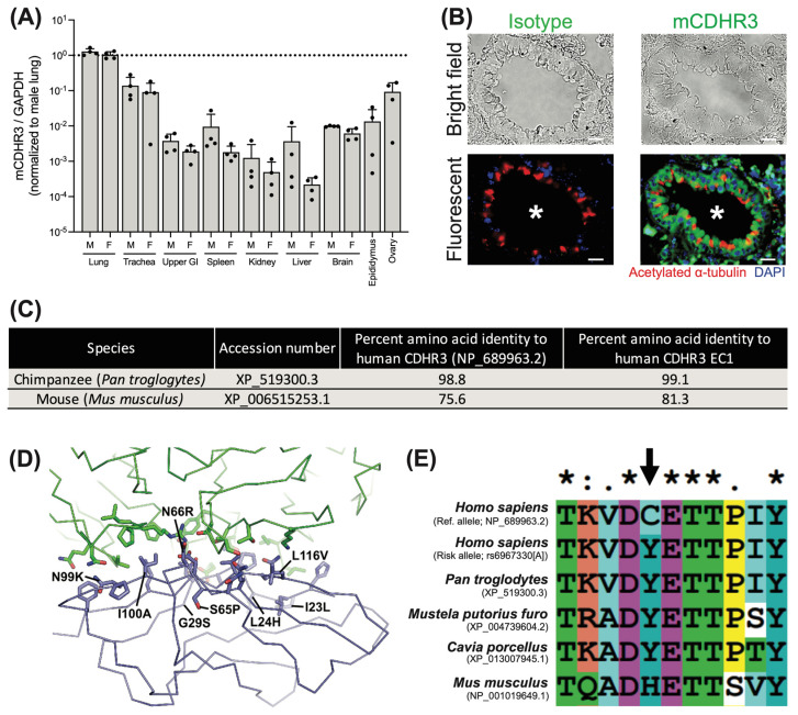
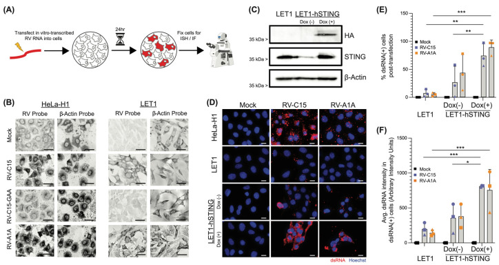
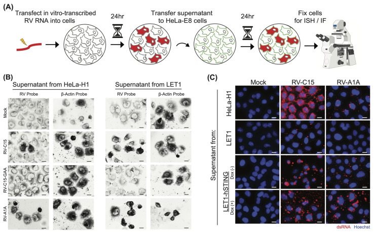
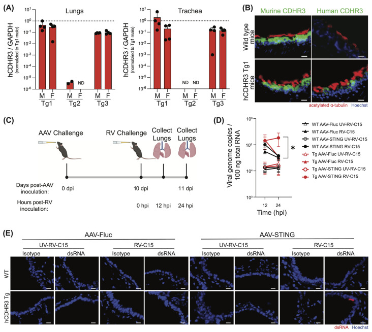
Rhinovirus C (RV-C) infects airway epithelial cells and is an important cause of acute respiratory disease in humans. To interrogate the mechanisms of RV-C-mediated disease, animal models are essential. Towards this, RV-C infection was recently reported in wild-type (WT) mice, yet, titers were not sustained. Therefore, the requirements for RV-C infection in mice remain unclear. Notably, prior work has implicated human cadherin-related family member 3 (CDHR3) and stimulator of interferon genes (STING) as essential host factors for virus uptake and replication, respectively. Here, we report that even though human (h) and murine (m) CDHR3 orthologs have similar tissue distribution, amino acid sequence homology is limited. Further, while RV-C can replicate in mouse lung epithelial type 1 (LET1) cells and produce infectious virus, we observed a significant increase in the frequency and intensity of dsRNA-positive cells following hSTING expression. Based on these findings, we sought to assess the impact of hCDHR3 and hSTING on RV-C infection in mice in vivo. Thus, we developed hCDHR3 transgenic mice, and utilized adeno-associated virus (AAV) to deliver hSTING to the murine airways. Subsequent challenge of these mice with RV-C15 revealed significantly higher titers 24 h post-infection in mice expressing both hCDHR3 and hSTING-compared to either WT mice, or mice with hCDHR3 or hSTING alone, indicating more efficient infection. Ultimately, this mouse model can be further engineered to establish a robust in vivo model, recapitulating viral dynamics and disease.

Keywords: CDHR3; STING; mouse model; rhinovirus C.


Conflict of interest statement

M.A.S. received funding from AstraZeneca PLC to support the studies reported here.

Figures

Figure 1
Figure 1
Murine CDHR3 is localized to similar tissues to human CDHR3, yet differs in several key aspects. (A) qPCR of murine CDHR3 expression relative to murine GAPDH by tissue across n = 4 male (M) and n = 4 female (F) mice. Mean +/− standard deviation. (B) Immunohistochemical detection of CDHR3 (green) in a BALB/c mouse lung tissue. Nuclei (DAPI; blue) and cilia (acetylated alpha tubulin; red). White asterisk indicates airway lumen. Scale bar = 20 µm. (C) Table detailing CDHR3 amino acid conservation in chimpanzees and mice. (D) The cryo-EM structure of RV-C15 (green) in complex with the human CDHR3 EC1 (blue) is shown as a Cα trace. The three RV-C15 capsid proteins that interact with EC1 (VP1, VP2, and VP3) are all shown as the same color. The side chains that comprise the RV-C15-EC1 interface are represented as sticks. Residues that differ from the mouse ortholog are labeled. (E) Homology of CDHR3 at position 529 (indicated by the black arrow), performed using ClustalX2.
Figure 2
Figure 2
Mouse cells replicate RV-C, and replication is significantly increased upon expression of human STING. (A) Experimental design schematic. HeLa-H1 cells or LET1 cells were transfected in parallel with in vitro-transcribed RV RNA or mock-transfected. (B) In situ hybridization (ISH) probes (dark) identify the presence of negative-sense RV RNA, indicative of replicating RV for RV-C15-transfected LET1 (right) and HeLa-H1 cells (positive control, left), but not in mock or replication-deficient (RV-C15-GAA) genome-transfected cells at 24 hpt. Scale bar = 20 µm. (C) Western blot shows inducible expression of hSTING in modified LET1 cells. (D) Immunofluorescence-mediated detection of dsRNA (red) in HeLa-H1 cells (positive control), LET1, and LET1-hSTING with or without induction after transfection with either RV-C15 or RV-A1A RNA (positive control), but not in mock-transfected wells, confirms RV-C15 replication in a mouse cell background. Staining was performed 24 hpt. Nuclei (Hoechst; blue). Scale bar = 20 µm. (E) CellProfiler quantification of the % dsRNA-positive cells as from panel (D). Each individual point (square, circle, or triangle) represents the mean from an independent experiment. In each experiment, >30 cells were assayed across n = 4 immunofluorescence images taken at predetermined locations within the culture. Error bars represent standard deviation of the mean across experiments. (F) CellProfiler quantification of the average dsRNA intensity in dsRNA-positive cells. Statistical analysis in panels (E,F) was performed using a Mann–Whitney U test (two-tailed; 0.95% confidence interval; * p < 0.05, ** p < 0.01, *** p < 0.001).
Figure 3
Figure 3
RV-C produced in mouse cells is infectious and capable of reinfection. (A) Experimental design schematic. HeLa-H1, LET1, or LET1-hSTING cells with or without induction of hSTING were transfected in parallel with in vitro-transcribed RV RNA or mock-transfected. At 24 hpt, supernatants were collected, filtered to remove any cellular material, and passaged to naïve HeLa-E8 cells. Infection of HeLa-E8 cells was confirmed by in situ hybridization or immunofluorescence staining. (B) In situ hybridization (ISH) probes (dark) identify the presence of negative-sense RV RNA, indicative of replicating RV in HeLa-E8 after inoculation with the supernatants described in (A), confirming infectious particle formation in LET1 cells. β-Actin ISH probes validate RNA integrity in the sample. Scale bar = 20 µm. (C) Immunofluorescence-mediated detection of dsRNA (red) in HeLa-E8 cells 24 hpi with supernatants harvested from HeLa-H1 (positive control), LET1, LET1-hSTING (with or without induction) cells previously transfected with either RV-C15 or RV-A1A RNA (positive control) but not mock. Nuclei (Hoechst; blue). Scale bar = 20 µm.
Figure 4
Figure 4
RV-C infection of transgenic mice expressing CDHR3 with the addition of human STING results in prolonged viral replication. (A) Human CDHR3 expression in the lungs and trachea of transgenic founder lines, Tg1, Tg2, and Tg3 by qPCR across n = 4 male (M) and n = 4 female (F) mice. Mean +/− standard deviation. ND = not detected. (B) Immunohistochemical detection of human and murine CDHR3 (green) in Tg1 mouse lung tissue compared to WT mice. Nuclei (Hoechst; blue) and cilia (acetylated alpha tubulin; red). Scale bar = 20 µm. (C) Experimental design schematic created with BioRender.com. Transgenic mice expressing human CDHR3 (Tg1) or WT mice were transduced with either human STING, or firefly luciferase (Fluc) via adeno-associated virus (AAV) by intranasal inoculation. On day 10 post-AAV transduction, mice were inoculated intranasally with RV-C15 or UV-inactivated RV-C15. Mice were then sacrificed, and total lungs were collected at 12 and 24 hpi. (D) qPCR detection of RV genome copy numbers at 12 and 24 hpi. Each point represents mean genome copy number from n = 11–14 mice assayed across three independent experiments. Black shapes represent WT mice and red shapes represent hCDHR3 Tg mice. Triangles represent mice receiving AAV-Fluc and circles represent mice receiving AAV-STING. Empty shapes represent UV-inactivated RV-C15 conditions while filled-in shapes represent mice receiving infectious RV-C15. Statistical analysis was performed using a Mann–Whitney U test (two-tailed; 0.95% confidence interval; * p < 0.05). (E) Immunohistochemical detection of dsRNA (J2; red) in paraffin-embedded mouse lung sections 24 hpi. Nuclei (Hoechst; blue). Scale bar = 20 μm.

References

    1. Arruda E., Pitkäranta A., Witek T.J., Doyle C.A., Hayden F.G. Frequency and natural history of rhinovirus infections in adults during autumn. J. Clin. Microbiol. 1997;35:2864–2868. doi: 10.1128/jcm.35.11.2864-2868.1997. - DOI - PMC - PubMed
    1. Lee W.M., Lemanske R.F., Evans M.D., Vang F., Pappas T., Gangnon R., Jackson D.J., Gern J.E. Human rhinovirus species and season of infection determine illness severity. Am. J. Respir. Crit. Care Med. 2012;186:886–891. doi: 10.1164/rccm.201202-0330OC. - DOI - PMC - PubMed
    1. Matsuno A.K., Gagliardi T.B., Paula F.E., Luna L.K.S., Jesus B.L.S., Stein R.T., Aragon D.C., Carlotti A.P.C.P., Arruda E. Human coronavirus alone or in co-infection with rhinovirus C is a risk factor for severe respiratory disease and admission to the pediatric intensive care unit: A one-year study in Southeast Brazil. PLoS ONE. 2019;14:e0217744. doi: 10.1371/journal.pone.0217744. - DOI - PMC - PubMed
    1. Ng K.T., Oong X.Y., Lim S.H., Chook J.B., Takebe Y., Chan Y.F., Chan K.G., Hanafi N.S., Pang Y.K., Kamarulzaman A., et al. Viral Load and Sequence Analysis Reveal the Symptom Severity, Diversity, and Transmission Clusters of Rhinovirus Infections. Clin. Infect. Dis. 2018;67:261–268. doi: 10.1093/cid/ciy063. - DOI - PubMed
    1. Corne J.M., Marshall C., Smith S., Schreiber J., Sanderson G., Holgate S.T., Johnston S.L. Frequency, severity, and duration of rhinovirus infections in asthmatic and non-asthmatic individuals: A longitudinal cohort study. Lancet. 2002;359:831–834. doi: 10.1016/S0140-6736(02)07953-9. - DOI - PubMed

Publication types

Supplementary concepts

LinkOut - more resources